Thorax Ct

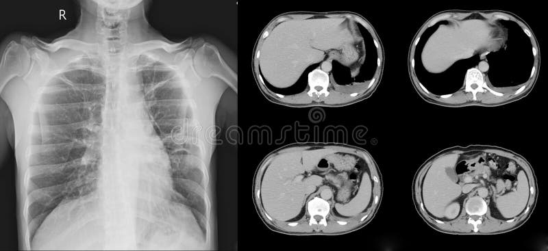
Ct Abdomen And Thorax Stock Photo Picture And Royalty Free Image Image

Image Ct Of Thorax Showing Anatomy Of Aorta And Pulmonary Artery Msd Manual Professional Edition
Q Tbn And9gcsolirq76ekc2ysm4lypsfn1xymluacqlh3prfyigfza7z27 Va Usqp Cau

Ct Abdomen And Thorax Stock Photo Picture And Royalty Free Image Image

Ct Scan Of Thorax And Abdomen Stock Photo Download Image Now Istock

Normal Ct Chest Radiology Case Radiopaedia Org
Each CT in cohort 2 was scored independently by two radiologists (BG and AJP) with 3 and 5 years of thoracic imaging experience, respectively Observers were blinded to all clinical information and the time points of the serial CTs.
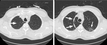
Thorax ct. Computed Tomography (CT)–Head, repeated with and without contrast material 4 mSv 16 months Computed Tomography (CT)–Spine 6 mSv 2 years CHEST Procedure Approximate effective radiation dose Comparable to natural background radiation for Computed Tomography (CT)–Chest 7 mSv 2 years Computed Tomography (CT)–Lung Cancer Screening 15 mSv 6. Diagnosis Thoracic aortic aneurysms are often found during routine medical tests, such as a chest Xray, CT scan or ultrasound of the heart, sometimes ordered for a different reason Your doctor will ask questions about your signs and symptoms, as well as your family's history of aneurysm or sudden death. Chest CT shows hazy opacities in both lungs predominantly peripheral in distribution (arrows) These are the typical features of the disease that have been reported with COVID19 infection (c, d) COVID19 crazy paving The patient is a 58yearold man who presented with lightheadedness.
Lung cancer screening with lowdose computed tomography for primary care providers external icon;. Chest (thorax) CT scans of the chest can look for problems with the heart, lungs, esophagus, the aorta, or even many of the tissues in the chest It can also includes parts of the upper abdomen and can pick up abnormalities of the liver, spleen and stomach. All visitation at Lifespan hospitals is temporarily suspended.
Highresolution CT thorax Highresolution CT scanning is very useful for assessing the architecture of the lung and does not involve iv contrast It acquires thin, noncontiguous slices, between 1 and 15 mm in thickness, sampling the parenchyma at 10–15 mm intervals. A computed tomography (CT) scan is a type of imaging test It uses Xrays and a computer to make detailed pictures of the inside of your chest These images are better than regular Xrays They can give more details about injuries or diseases of the chest organs. The noncontrast CT chest is a commonly performed diagnostic examination It is often performed to evaluate conditions impacting the lungs or preferenced over a contrast enhanced scan when iodinated contrast is contraindicated NB This article.
It appears a thorax scan is the same as a chest scan but includes the upper abdomen But the reason for each type of scan is basically the same and in your case I think they are The radiologist will be supplied with info from your doctors regarding what items to take special note of to look for. CT scan upper back How the Test is. In this module of the animal atlas vetAnatomy is displayed the crosssectional labeled anatomy canine thorax on a Computed Tomography (CT) and on 3D images of the thorax of the dog CT images are available in 3 different planes (transverse, sagittal and dorsal) with two kinds of contrast (bones/lungs and soft tissues/mediastinum/vessels).
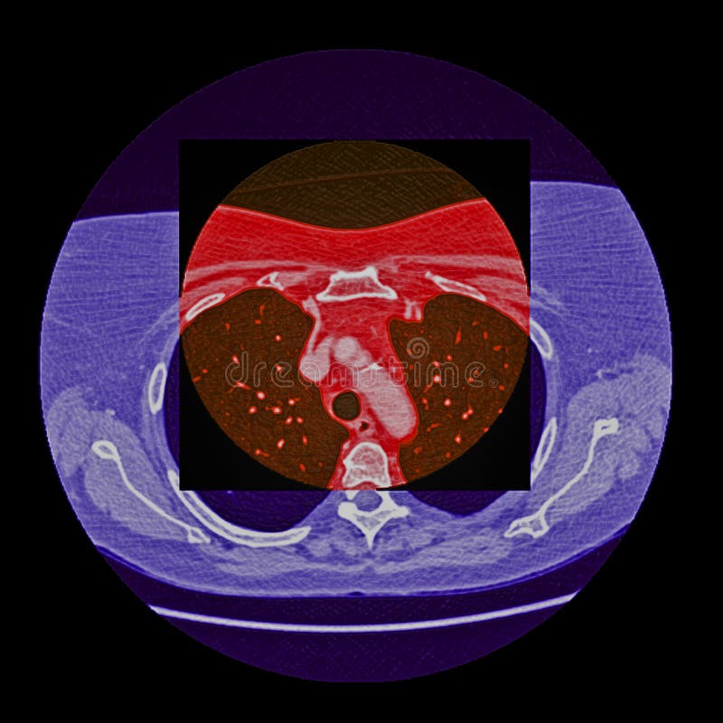
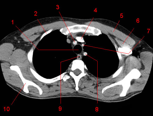
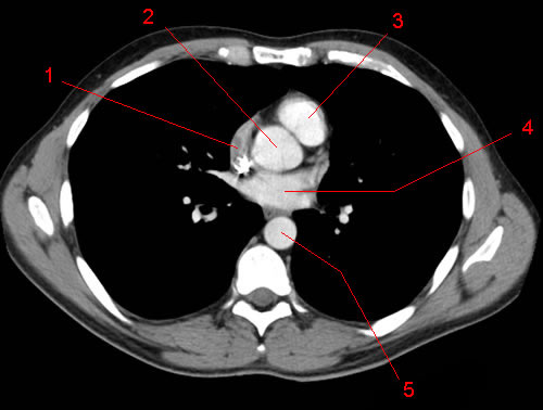
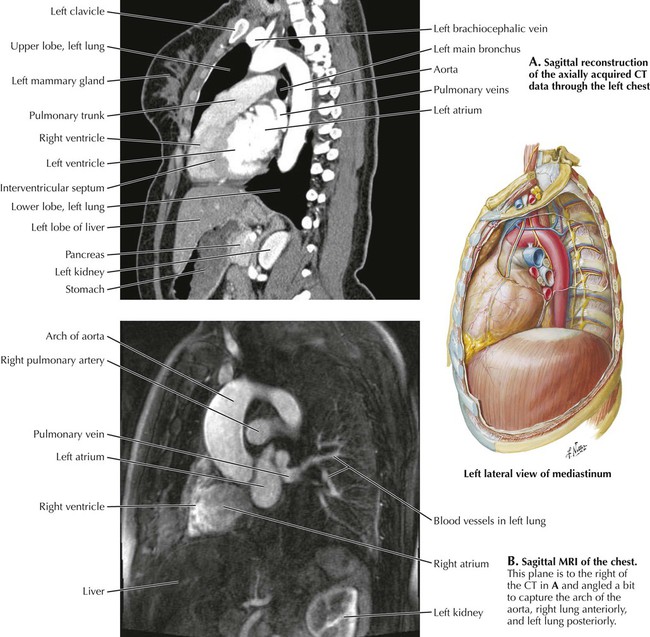
CT Chest Imaging Request For NONURGENT requests, please fax this completed document along with medical records, imaging, tests, etc If there are any inconsistencies with the medical office records, please elaborate in the comment section. Computed Tomography (CT or CAT) Scan of the Chest (Chest CT Scan, Thoracic CT Scan, CT of the Thorax). CT Anatomy of the chest, axial reconstruction 1, Lung (right side) 2, Ventricle (right side) 3, Ventricle (left side) 4, Lung (left side) 5, Descending thoracic aorta.
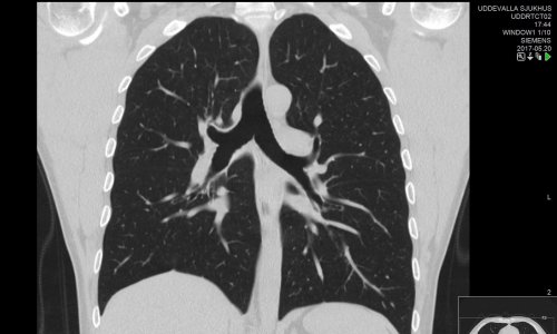
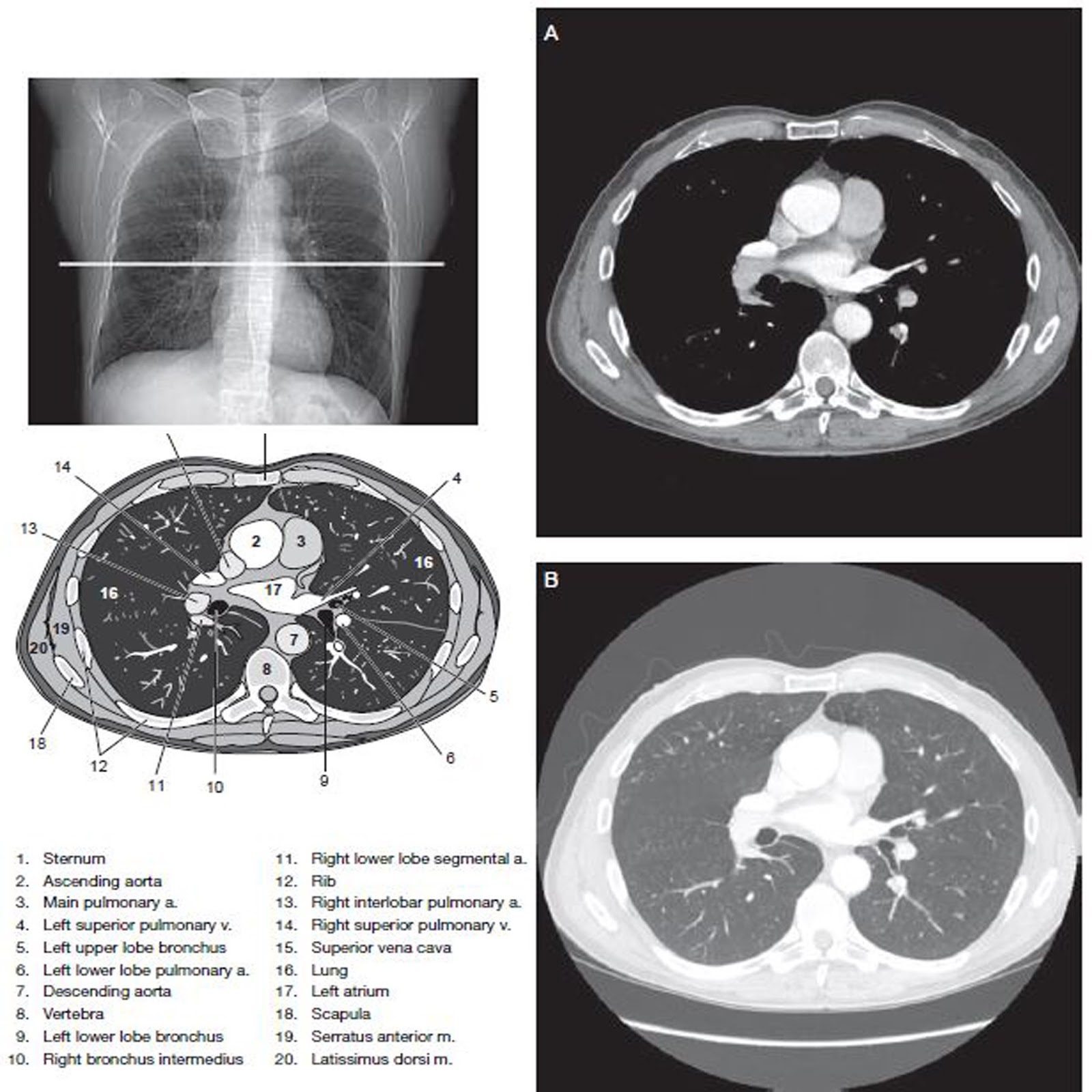
Normal CT of the chest performed with intravenous contrast Scan during the arterial phase. CT Scan of the Chest What is a CT scan of the chest?. Lung cancer kills an estimated 35 000 people in the UK every year Despite the improvements in treating latestage disease, lung cancer outcomes have changed little in the last 40 years Lowdose CT (LDCT) screening for lung cancer reduces lung cancer mortality by %–24% and allcause mortality by 7%1 2 Lung cancer screening (LCS) however remains contentious, particularly how to implement.
Chest CT is valuable to detect both alternative diagnoses and complications of COVID19 (acute respiratory distress syndrome, pulmonary embolism, and heart failure), while its role for prognostication requires further investigation. COVID19 pneumonia imaging and specific respiratory complications for consideration In typical cases of COVID19 pneumonia, the chest Xray (CXR) shows multiple bilateral peripheral opacities ()In some patients, the morphological pattern of lung disease on CT scan with regions of groundglass opacification and consolidation, which variably comprise foci of oedema, organising pneumonia and. Ct thorax w/o dye Ct thorax w/dye Ct thorax w/o & w/dye Please refer to the CMS website for the ICD10 Codes that Support Medical Necessity Documentation Requirements Documentation must be legible, relevant and sufficient to justify the services billed This documentation must be made available to the A/B MAC upon request.
A computed tomography (CT) scan is a type of imaging test It uses Xrays and a computer to make detailed pictures of the inside of your chest These images are better than regular Xrays They can give more details about injuries or diseases of the chest organs. The NEXUS Chest CT research team should be applauded for rigorously addressing a common issue of Chest CT utilization for blunt trauma patients in the era of panCT’ing Developing a 2tier decision instrument, the Chest CTMajor and Chest CTAll, helps institutions address whether they wish to use a more risktolerant or riskaverse approach. Start to finish search pattern of a noncontrast chest ct / thorax ct For beginner levelOrganizationIntro 0000Lung Parenchyma 136Pleura 338Airway.
Computed tomography scan thoracic spine;. Mazzone PJ, Silvestri GA, Patel S, Kanne JP, Kinsinger LS, Wiener RS, Soo Hoo G, Detterbeck FC Screening for lung cancer CHEST guideline and expert panel report external icon Chest 18;153(4)954–985 DOI /jchest. CT scan is a type of imaging test It uses Xray and computer technology to make detailed pictures of the organs and structures inside your chest These images are more detailed than regular Xrays They can give more information about injuries or diseases of the chest organs.
Each CT in cohort 2 was scored independently by two radiologists (BG and AJP) with 3 and 5 years of thoracic imaging experience, respectively Observers were blinded to all clinical information and the time points of the serial CTs. Introduction Contiguous CT of the Thorax, Abdomen, and Pelvis A normal CT of the chest, abdomen and pelvis, consisting of 130 3mm thick axial sections, has been labelled for your review and self learning It begins on the next page, with the first image of the chest, and continues through the pelvis. Ct thorax w/o dye Ct thorax w/dye Ct thorax w/o & w/dye Please refer to the CMS website for the ICD10 Codes that Support Medical Necessity Documentation Requirements Documentation must be legible, relevant and sufficient to justify the services billed This documentation must be made available to the A/B MAC upon request.
A CT scan (also called a CAT scan) is a noninvasive, painless medical test that helps physicians diagnose and treat medical conditions CT scans allow physicians to rapidly create detailed pictures of the body allowing them to more easily diagnose problems such as cancers, cardiovascular disease, infectious disease, trauma and musculoskeletal disorders. · CT of the Thorax is indicated for assessing the appropriateness and feasibility of percutaneous procedures such as biopsy and pleural/parenchymal drainage CT of the thorax is also indicated for following for sequalae of, and response to treatment of these procedures. A CT scan (also called a CAT scan) is a noninvasive, painless medical test that helps physicians diagnose and treat medical conditions CT scans allow physicians to rapidly create detailed pictures of the body allowing them to more easily diagnose problems such as cancers, cardiovascular disease, infectious disease, trauma and musculoskeletal disorders.
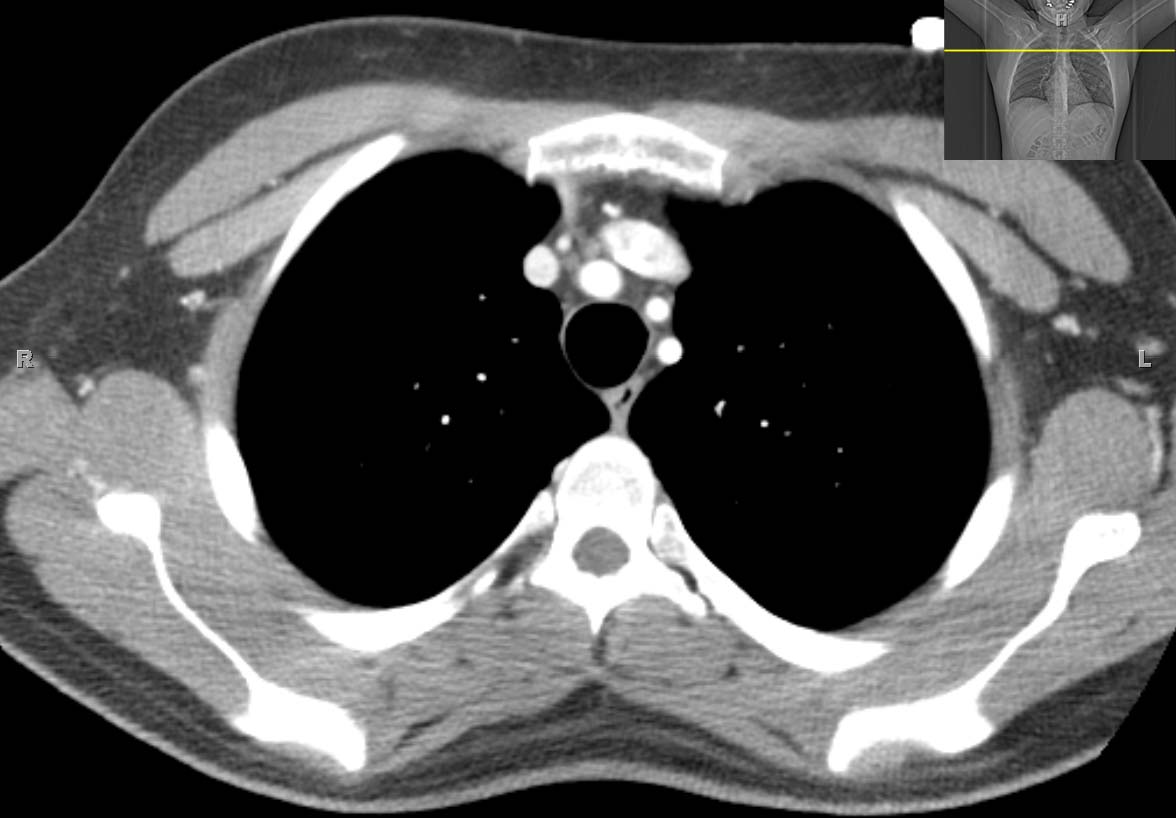
There are different ways to perform a chest CT, depending on what your doctor is looking for A "routine" CT will show lungs, mediastinum, aorta and great vessels, heart, ribs, upper abdomen, thoracic spine. Computed axial tomography scan thoracic spine;. CT Anatomy of the chest, axial reconstruction 1, Lung (right side) 2, Ventricle (right side) 3, Ventricle (left side) 4, Lung (left side) 5, Descending thoracic aorta.
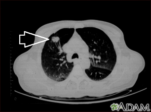
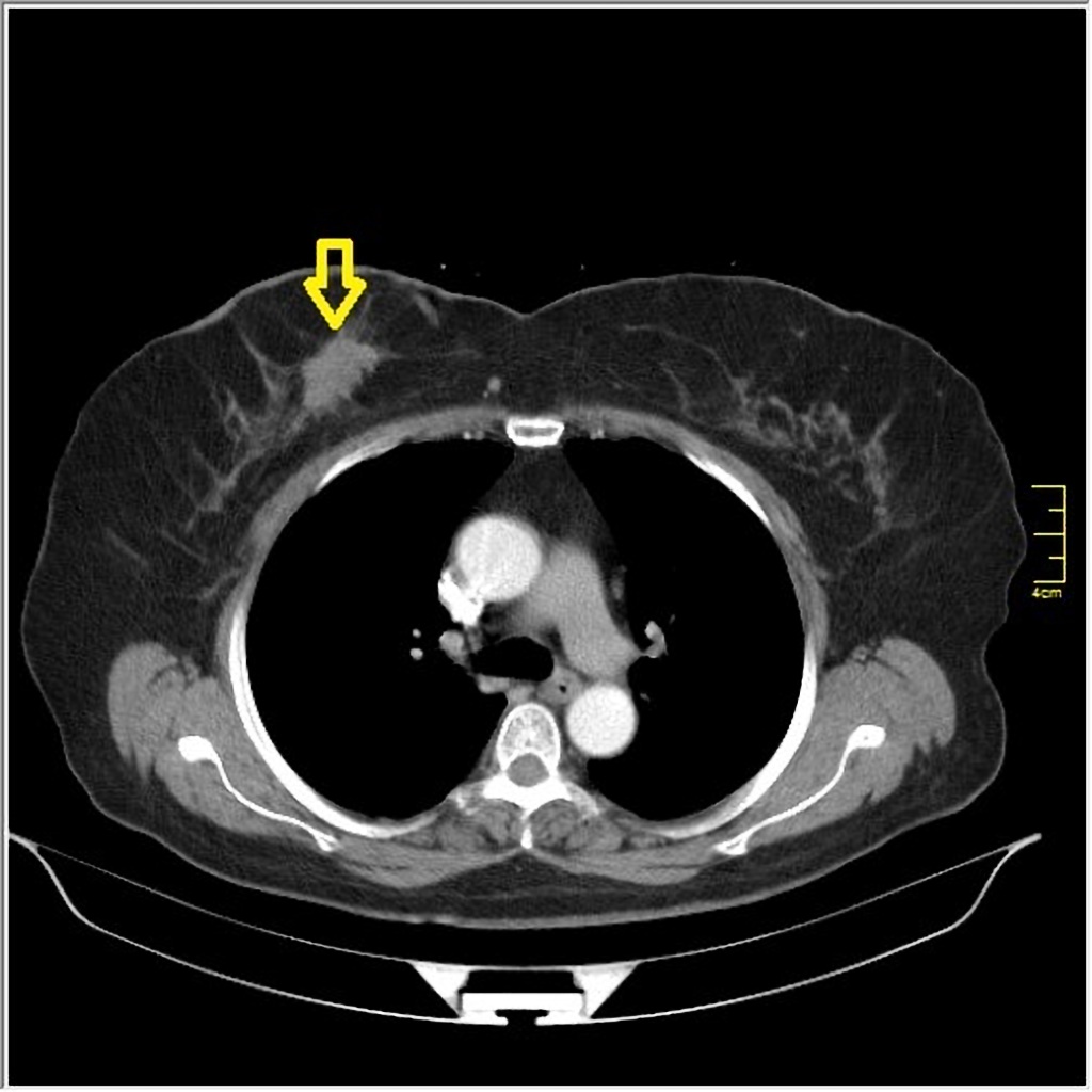
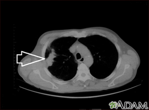
A computed tomography (CT) scan of the thoracic spine is an imaging method This uses xrays to rapidly create detailed pictures of the middle back (thoracic spine) Alternative Names CAT scan thoracic spine;. A chest CT (computed tomography) scan is an imaging method that uses xrays to create crosssectional pictures of the chest and upper abdomen This is a CT scan of the upper chest showing a mass in the right lung (seen on the left side of the picture) A CT scan showing a mass in right lower chest near the heart (left side of photograph). All visitation at Lifespan hospitals is temporarily suspended.
A computed tomography (CT) scan of the thoracic spine is an imaging method This uses xrays to rapidly create detailed pictures of the middle back (thoracic spine) Alternative Names CAT scan thoracic spine;. Anatomy of the thorax (lungs and mediastinum) (CT) interactive atlas of human anatomy using crosssectional imaging We created an anatomy atlas of the chest and the mediastinum which is an interactive tool for studying the crosssectional anatomy of the normal thorax based on an enhanced multidetector computed tomography with helical. CT Scan of the Chest What is a CT scan of the chest?.
CT scan upper back How the Test is. There are different ways to perform a chest CT, depending on what your doctor is looking for A "routine" CT will show lungs, mediastinum, aorta and great vessels, heart, ribs, upper abdomen, thoracic spine. Start to finish search pattern of a noncontrast chest ct / thorax ct For beginner levelOrganizationIntro 0000Lung Parenchyma 136Pleura 338Airway.
The noncontrast CT chest is a commonly performed diagnostic examination It is often performed to evaluate conditions impacting the lungs or preferenced over a contrast enhanced scan when iodinated contrast is contraindicated NB This article. Ct thorax w/o dye Ct thorax w/dye Ct thorax w/o & w/dye Please refer to the CMS website for the ICD10 Codes that Support Medical Necessity Documentation Requirements Documentation must be legible, relevant and sufficient to justify the services billed This documentation must be made available to the A/B MAC upon request. Normal CT of the chest performed with intravenous contrast Scan during the arterial phase.
CT Scan of the Chest What is a CT scan of the chest?. Computed axial tomography scan thoracic spine;. It appears a thorax scan is the same as a chest scan but includes the upper abdomen But the reason for each type of scan is basically the same and in your case I think they are The radiologist will be supplied with info from your doctors regarding what items to take special note of to look for.
CT scan upper back How the Test is. Lung cancer screening with lowdose computed tomography for primary care providers external icon;. These scans also encompass the diaphragm and the upper abdomen The distance (or volume) needed to cover the thorax (usually 25–30 cm) is determined by a preliminary projection scan (eg, a.
There are different ways to perform a chest CT, depending on what your doctor is looking for A "routine" CT will show lungs, mediastinum, aorta and great vessels, heart, ribs, upper abdomen, thoracic spine. "on a ct thorax what is dependent compressive atelectasis confirmed with prone and supine imaging?. CE CT shaded surface display of the right lung from scan of the thorax • The left lung is composed of two lobes (superior and inferior) separated by an oblique (major) fissure Only gold members can continue reading.
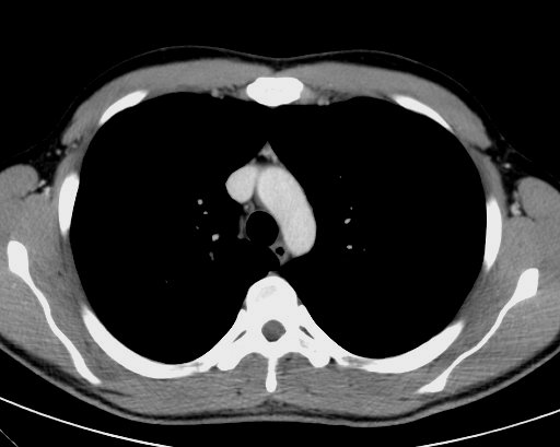
An overview of the anatomy visible in a transverse computed axial tomographical image of the thorax (and part of the abdomen) performed with intravenous cont. Find protocols for performing Chest CT scans at Rhode Island Hospital, also welcoming Massachusetts patients Coronavirus COVID19 Information Information for patients who have a scheduled test, appointment, surgery or telehealth visit;. The chest CT has high sensitivity for detecting viral pneumonia, even before clinical symptoms develop Further, the chest CT varies with disease course and severity as touched upon below Chest CT Abnormalities before Symptoms Shi and colleagues retrospectively reviewed the chest CT findings of 81 patients with confirmed COVID19 Of note.
Computed axial tomography scan thoracic spine;. Dr said my scan was normal" Answered by Dr Paxton Daniel Atelectasis Is lesswell inflated lung It can look like scarring or. Computed tomography (CT or CAT scan) of the chest is a special noninvasive Xray scan used to detect and diagnose health problems in the thorax (chest area) This scan can be used to evaluate symptoms such as shortness of breath, chest pain, unexplained cough, fever, and other chest symptoms.
A computed tomography (CT) scan is a type of imaging test It uses Xrays and a computer to make detailed pictures of the inside of your chest These images are better than regular Xrays They can give more details about injuries or diseases of the chest organs. Mazzone PJ, Silvestri GA, Patel S, Kanne JP, Kinsinger LS, Wiener RS, Soo Hoo G, Detterbeck FC Screening for lung cancer CHEST guideline and expert panel report external icon Chest 18;153(4)954–985 DOI /jchest. A computed tomography (CT) scan of the thoracic spine is an imaging method This uses xrays to rapidly create detailed pictures of the middle back (thoracic spine) Alternative Names CAT scan thoracic spine;.
Computed tomography scan thoracic spine;. Computed Tomography or CT Scan is one of the advanced Xray procedures Different from the usual Xray, CT scan employs multiple Xray beams and creates a detailed, 3Dlike image of the body parts CT scan of chest—also CT scan thorax—focuses on lungs and is designed for identification of various lungrelated disorders. Chest CT is usually performed from a level just above the lung apices (near the suprasternal notch) to the level of the posterior costophrenic angles;.
A thoracic CT scan, or computer tomography scan, provides a series of xrays of organs and structures in the chest and upper abdominal region Detecting internal bleeding or fluid filled areas, evaluating a chest injury, or assessing the position and size of organs are some of the reasons for having a CT scan. Computed tomography scan thoracic spine;. Computed Tomography or CT Scan is one of the advanced Xray procedures Different from the usual Xray, CT scan employs multiple Xray beams and creates a detailed, 3Dlike image of the body parts CT scan of chest—also CT scan thorax—focuses on lungs and is designed for identification of various lungrelated disorders.
Find protocols for performing Chest CT scans at Rhode Island Hospital, also welcoming Massachusetts patients Coronavirus COVID19 Information Information for patients who have a scheduled test, appointment, surgery or telehealth visit;.

Untitled Document

Chest Ct Scan St Elizabeth S Medical Center Steward Family Hospital Brighton Ma
Thorax Ct Photos Free Royalty Free Stock Photos From Dreamstime

Thorax Mediastinum Heart And Great Vessels Tracheal Dimensions By Ct Radiology Key

What Are Chest Or Thorax Ct Scans Two Views

Ct Scan Of Thorax And Abdomen Stock Photo Download Image Now Istock

Lung Cancer Thorax Ct Image Stock Photo Download Image Now Istock

Thorax Ct Image Stock Photo Download Image Now Istock

Registration Of Thorax Abdomen 3d Ct Scans For Oncological Follow Up Download Scientific Diagram
Thorax Ct Anatomy Anatomy Drawing Diagram
Thorax Ct Photos Free Royalty Free Stock Photos From Dreamstime

Thorax Radiologic Anatomy

Ct Chest Anatomy Axial Anatomy Of The Thorax Studykorner

Ct Scan Of The Thorax Ct Scan Of The Thorax Showing A Open I
Ct Thorax Transvers 2 Anatomytool
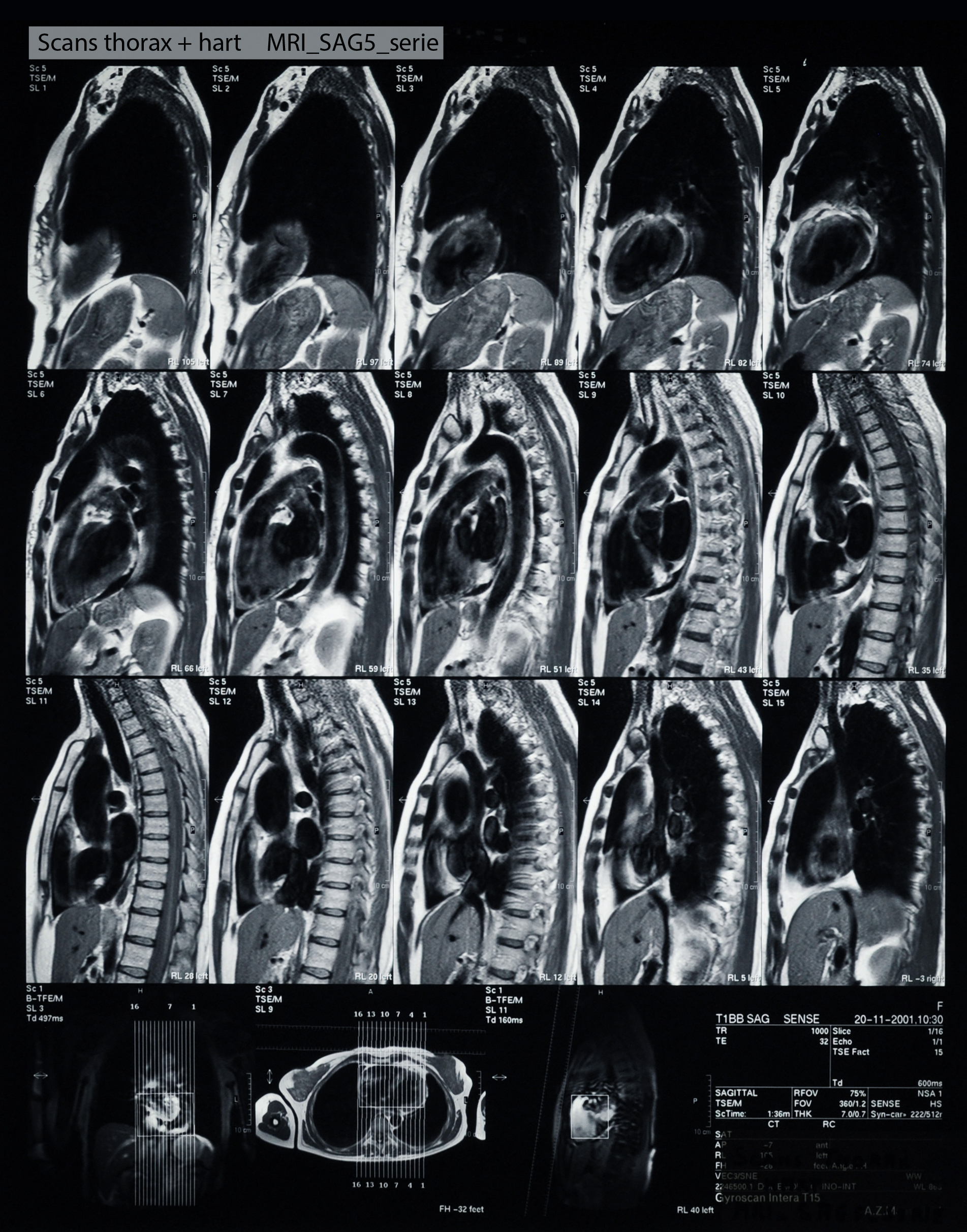
Ct Thorax Sagital Anatomytool

19 Ncov Milchglastrubungen Und Konsolidierungen Im Thorax Ct

Radiology Basics Chest Anatomy

Normal Cta Thorax Ecg Gated Radiology Case Radiopaedia Org
Public Quality Measure Report Thorax Ct Use Of Contrast Material

Axial Thorax Ct Scan Showing A Hypodense Well Defined Mass With Download Scientific Diagram

Thoracic Ct Information Mount Sinai New York

Normal Chest Ct Radiology Case Radiopaedia Org

Idcm Infectious Diseases And Clinical Microbiology
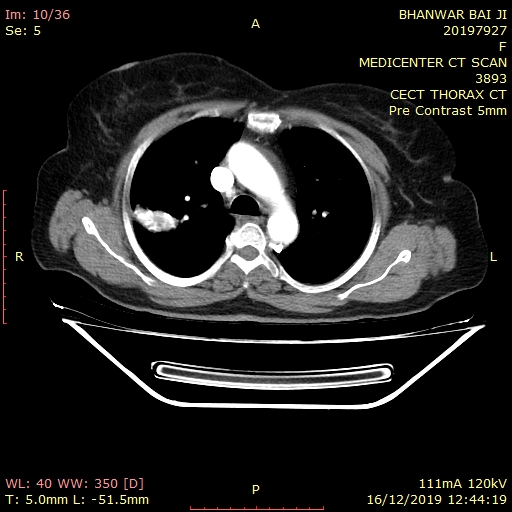
Diagnostic Centre Best Diagnostic Centre In Udaipur

Axial Ct Slice Shows The Measurement Of Thorax Ap And Lateral Diameters Download Scientific Diagram

Thorax Ct Ihr Online Portal Fur Mitglieder Und Interessierte

Abbildung 4a B Thorax Ct

Ct Procedure Of Thorax Ct Chest

What Are Chest Or Thorax Ct Scans Two Views
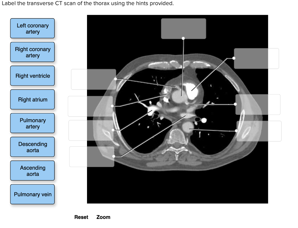
Solved Label The Transverse Ct Scan Of The Thorax Using T Chegg Com

File Thorax Ct 09 N Jpg Wikimedia Commons

Lung Cancer Thorax Ct Image Stock Photo Download Image Now Istock

Figure1 Findings Of Abdominal Chest Thorax On Ct And Chest X Ray A Download Scientific Diagram

Ct Scan Of Thorax And Abdomen Stock Photo Download Image Now Istock

Ct Abdomen And Thorax Stock Photo Picture And Royalty Free Image Image
Atlas Of Ct Anatomy Of The Chest W Radiology

High Resolution Computed Tomography Wikipedia
Q Tbn And9gcqmvycwc1oqbtu7i1nq7ji8praufjjbdd0mjbfzqvc Usqp Cau

Post Contrast Axial Ct Scan Through The Thorax Download Scientific Diagram

Computertomographie Des Thoraxbereiches Lunge Ct Mrt Institut Berlin

Ct Head Thorax Ct Vet Coach Llc
3

Anatomy Of The Thorax Ct

Thorax Of The Dog Cross Sectional Anatomy On Computed Tomography Ct

Which Parts Of The Human Body Does A Thoracic Ct Scan Cover Can Any Quora User Provide A 2d 3d Image Of The Human Body Showing The Area Of Coverage Of A Thoracic

Thorax Ct Scan High Res Stock Photo Getty Images
Breast Cancer On Thorax Ct Radiology Case Radiopaedia Org
:background_color(FFFFFF):format(jpeg)/images/library/12302/ct-axial-t3-level_english.jpg)
Medical Imaging And Radiological Anatomy X Ray Ct Mri Kenhub

What Are Chest Or Thorax Ct Scans Two Views

Cat Scan Ct Chest

What Are Chest Or Thorax Ct Scans Two Views
Thorax Ct Bei Covid 19 Patient Worauf Zeigen Die Pfeile Kardiologie Org

Apical Lung Herniation Thorax
Figure 2 Unklarer Thoraxschmerz Bei Einem Jahrigen Springerlink
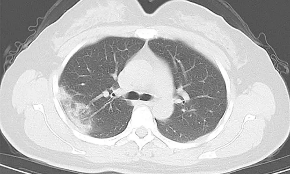
Ct Thorax Scan Has Over 90 Covid Test Accuracy

Anatomy Of The Thorax Ct

What Are Chest Or Thorax Ct Scans Two Views
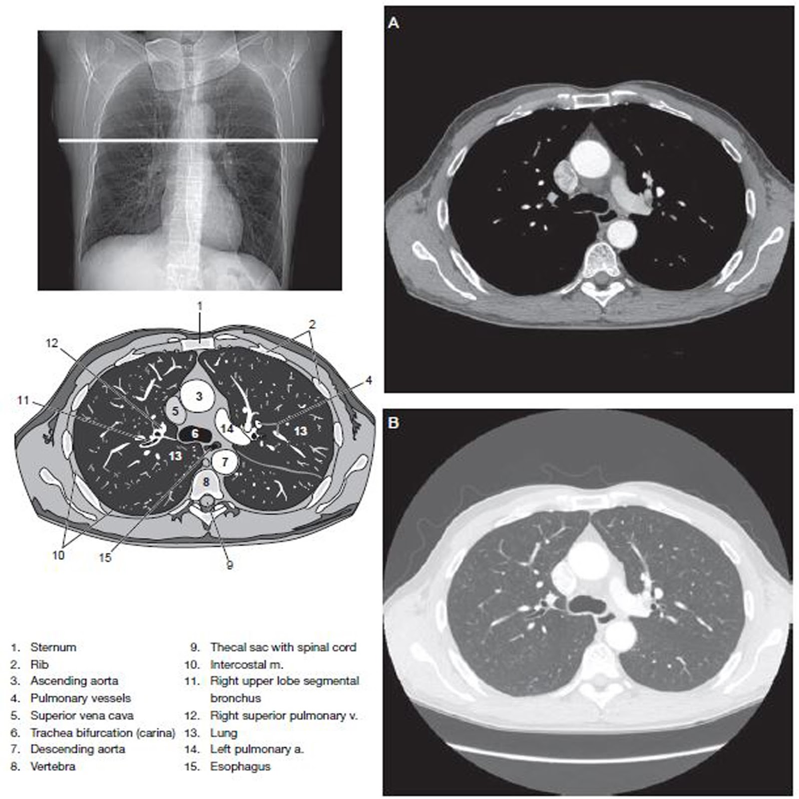
Chest Ct Scan Imaging Radtechonduty

Chest Ct Scan Imaging Radtechonduty

A Initial Ct Scan Of The Thorax Showing Minimal Lung Abnormality B Download Scientific Diagram
%20axial%20image%2031.jpg)
Chest Anatomy Mri Chest Thorax Axial Anatomy Free Cross Sectional Anatomy

Abbildung 2b Thorax Ct

High Resolution Ct Thorax

Thorax Ct Scan With The Tumor But With No Mediastinal Lymphadenopathy Download Scientific Diagram

Part2 Ct Thorax Normal Anatomy Youtube

Normal Thorax Ct Youtube

Radiology Basics Chest Anatomy

Asthma High Resolution Ct Scan Of The Thorax Demo Radiology Radiography Medical Knowledge

Anatomy Of The Thorax Ct

Anatomy Of The Thorax Ct

Ct Thorax

Cross Sectional And Imaging Anatomy Of The Thorax Youtube
%20axial%20image%2037.jpg)
Chest Anatomy Mri Chest Thorax Axial Anatomy Free Cross Sectional Anatomy

Ct Scan Of Thorax And Abdomen Stock Photo Download Image Now Istock

Thorax Ct Scan Showed Centrilobular Nodules With Lobulated Contours And Download Scientific Diagram
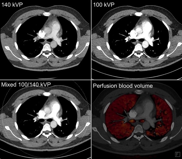
Dual Energy Ct Thorax Technique And Application Radiology Case Radiopaedia Org

Images Learn Nlst The Cancer Data Access System

Computertomographie Des Thoraxbereiches Lunge Ct Mrt Institut Berlin
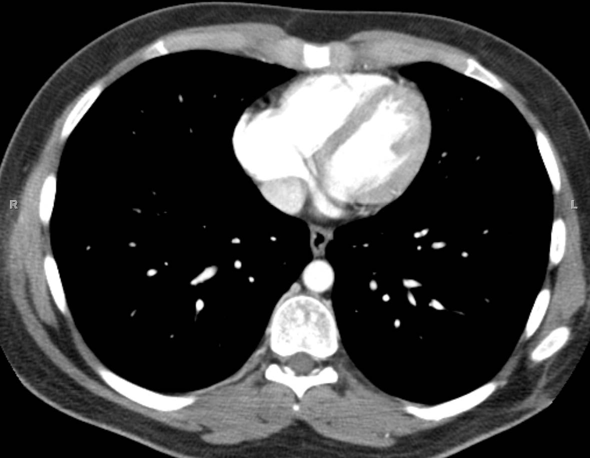
Low Cardiac Level Computed Tomograph Axial Thorax Ct
Ct Outperforms Lab Diagnosis For Coronavirus Infection

Fig A3 4 Ct Anatomy Of The Thorax Level Of T6 Oxford Medicine Online
Supracardiac Level Computed Tomograph Axial Thorax Ct

Axial Anatomy Of The Thorax Radiology Radiology Student Radiology Imaging
Atlas Of Ct Anatomy Of The Chest W Radiology

Abb 1 8 Ct Axiale Schichtfuhrung Uber Den Thorax Mit Kontrastmittel Download Scientific Diagram
Imaging Anatomy Interactive Pacs Like Atlas Of Radiological Anatomy
Thoracic Ct Information Mount Sinai New York

Epos Trade

Cross Sectional Ct Anatomy Thorax Flashcards Quizlet

Anatomy Of A Transverse Ct Of The Thorax Youtube
3

Ct Scan Of The Thorax Cect Showing A Large Mass Of Heterogeneous Download Scientific Diagram

Diagnostic Centre Best Diagnostic Centre In Udaipur

Neck And Thorax Ct Scan The Pre Treatment Figures Show A Large Mass Download Scientific Diagram
Thorax Radiology Key

Thorax Ct Showing Multiple Small Excavated Nodules Scat Open I
Chest Ct Scan Imaging Radtechonduty


