Arteria Carotis Interna Segments
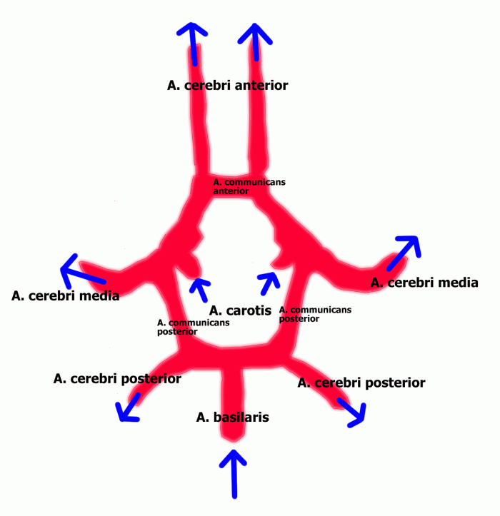
Arterien Des Gehirns
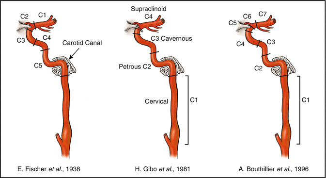
Essential Neurovascular Anatomy Springerlink
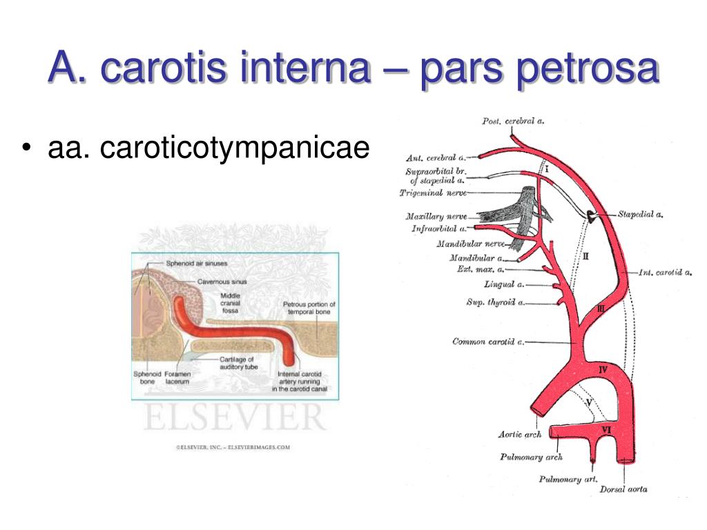
Ppt Arterial System Systema Arteriarum Powerpoint Presentation Free Download Id
Q Tbn And9gctk7ur7ul Fvt7zlnwnfovv Gniep 26i4nopgn1sqxo8gcvo0n Usqp Cau

Internal Carotid Artery Parts Course Relations Of Cervical Part Youtube

Internal Carotid Artery Radiology Reference Article Radiopaedia Org
M2 is the insular part (pars insularis), the segment running in the lateral cerebral ( sylvian) fissure and serving the insular cortex;.
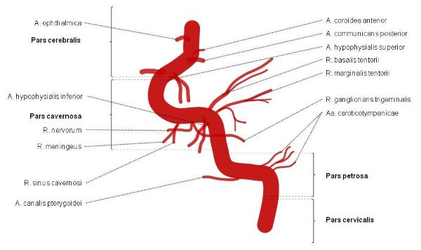
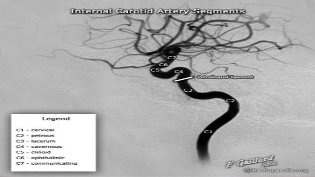
Arteria carotis interna segments. C2 petrous (horizontal) segment caroticotympanic artery;. The segments of the internal carotid artery are as follows Cervical segment, or C1, identical to the commonly used Cervical portion Petrous segment, or C2 Lacerum segment, or C3 C2 and C3 compose the commonly termed Petrous portion Cavernous segment, or C4, almost identical to the commonly used Cavernous portion. Carotis Segments Images from MR Angiography (MRA) with Time of Flight (ToF) source sequence on the left and reconstructed Maximum Intensity Projection (MIP) showing stenoses in the Internal Carotid Artery (ICA) This patient has a stent in the distal carotid artery that beginns in petrous and ends in cavernous segment.
Die Arteria carotis interna (oder innere Halsschlagader, Abkürzung ACI) gehört zu den hirnversorgenden SchlagadernBeim Menschen und einigen anderen Säugetieren versorgt sie neben dem Gehirn auch das AugeDer Ast wird Arteria ophthalmica genannt Diese Seite wurde zuletzt am 3 Mai um 1221 Uhr bearbeitet. Ziel der Arbeit ist die Darstellung vaskulitisch bedingter Veränderungen der Arteria carotis interna infolge einer Riesenzellarteriitis (Arteriitis temporalis) Bei zwei von neun zwischen 1996 und 1998 konsekutiv, behandelten Patienten mit histologisch gesicherter Arteriitis temporalis fanden sich angiographisch für eine Vaskůlitis typische Veränderungen („vascular narrowings”) in. Arteria carotis interna kan motta blodforsyning via en viktig kollateral blodforsyning til hjernen, den cerebrale arterielle sirkel, bedre kjent som Willis' arterielle sirkel Grener Følgende liste navngir forgreningene til arteria carotis interna, sortert etter segment C1 Grener fra cervicalsegmentet – ingen.
A short video tutorial demonstrating the route of the internal carotid artery and its relationship to the cavernous sinus A 3D anatomical model is used alon. The authors' classification has the following seven segments C1, cervical;. The four segments into which the posterior cerebral artery is divided;.
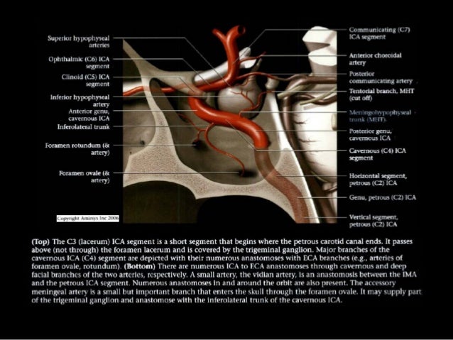
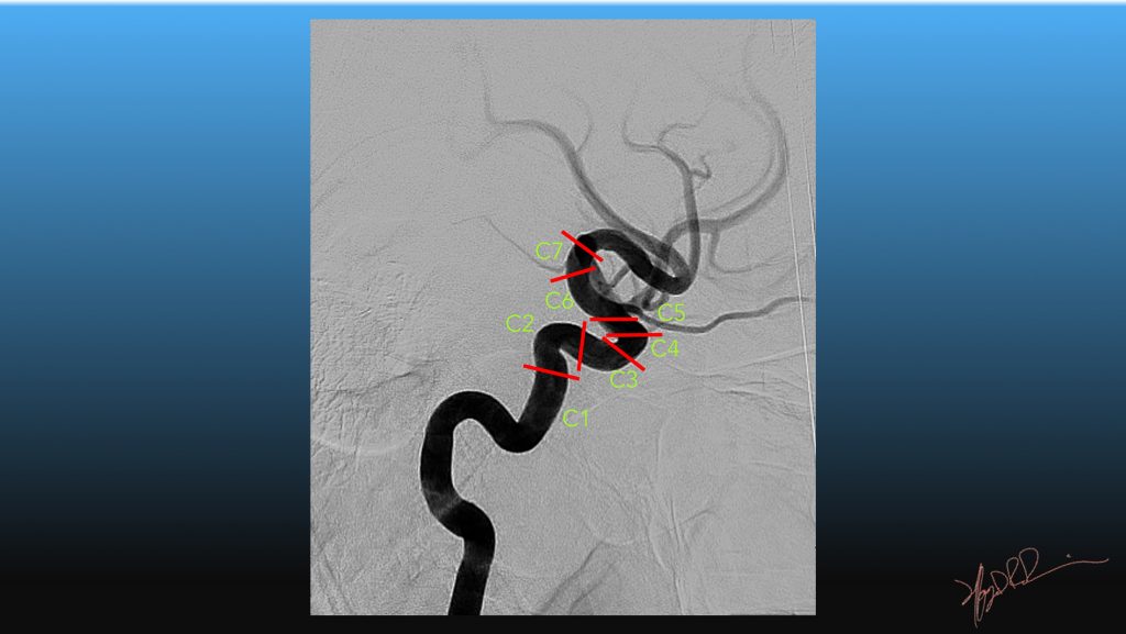
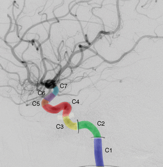
C4 cavernous segment meningohypophyseal trunk;. Ophthalmic (supraclinoid) segment C6;. Bouthillier classification of internal carotid artery segments Dr Francis Deng and Assoc Prof Frank Gaillard et al Alain Bouthillier et al described a seven segment internal carotid artery (ICA) classification system in 1996 1 It remains the most widely used system for describing ICA segments A helpful mnemonic for remembering ICA segments is.
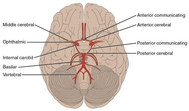
One of a pair found on each side of the neck, the internal carotid artery branches off from the common carotid artery and works its way up into the cranium Its path places it right alongside brain regions associated with visual and sensory processing and, at its end, it splits into the two cerebral arteries. P2 is the postcommunicating part (pars postcommunicalis), which extends from there to the temporal branches;. Communicating (terminal) segment C7.
7 segments of Internal Carotid Artery We have already discussed a mnemonic to remember the course of Internal Carotid Artery with the help of 2 horizontal “S” under the topic of Circle of Willis C1 – Cervical segment C2 – Petrous (horizontal) segment C3 – Lacerum segment C4 – Cavernous segment C5 – Clinoid segment. Origin, terminal Medical dictionary. Arteria carotis interna est arteria, quae cerebrum et (per arteriam ophthalmicam) oculum rigat Ad usum clinicum ab neurochirurgo BOUTHILLIER cogitata est arteria carotis interna in segmenta anatomica septem, C1C7 Interdum arteria carotis interna cum arteria carotide externa per fistulam cavernosam carotidem communicat, cum symptomate exophthalmo pulsatili et chemosi (tumescentia tunicae.
OBJECTIVE To reconcile various internal carotid artery (ICA) nomenclatures for transcranial and endoscopicendonasal perspectives, we reexamined the transition between lacerum (C3) and cavernous (C4) segments using a C1C7 segments schema In this cadaveric study, we obtained a 360°circumferential view integrating histological. The four segments into which the middle cerebral artery is divided, M1â €“ M4 in clinical terminology The M1 segment is the sphenoid part (pars sphenoidalis), the horizontal portion of the vessel from the termination of the internal carotid artery to the point of branching at the limen insulae;. At its origin, this artery is closer to the skin and more medial than the internal carotid, and is situated within the carotid triangle Development edit In children, the external carotid artery is somewhat smaller than the internal carotid ;.
Arteria carotis interna kan motta blodforsyning via en viktig kollateral blodforsyning til hjernen, den cerebrale arterielle sirkel, bedre kjent som Willis' arterielle sirkel Grener Følgende liste navngir forgreningene til arteria carotis interna, sortert etter segment C1 Grener fra cervicalsegmentet – ingen. A short video tutorial demonstrating the route of the internal carotid artery and its relationship to the cavernous sinus A 3D anatomical model is used alon. C1 Cervical segment C1 Cervical segment Level of 6th cervical vertebrae still at level of common carotid but relationships are similar to C2 Petrous segment The petrous segment, or C2, of the internal carotid, is that which is inside the petrous part of the C4 Cavernous segment Oblique.
M3 is the opercular part, the segment emerging from the. M2 is the insular part (pars insularis), the segment running in the lateral cerebral ( sylvian) fissure and serving the insular cortex;. Die Arteria carotis interna ist auch als innere Halsschlagader bekannt und versorgt Anteile des Gehirns mit arteriellem Blut Gemeinsam mit der Arteria carotis externa geht sie aus der Arteria carotis communis hervor Die innere Halsschlagader ist für Arteriosklerose sowie kleinere Aneurysmen besonders anfällig.
The internal carotid artery segments, according to the Bouthillier classification, can be recalled by the following mnemonic C'mon Please Learn Carotid Clinical Organizing Classification;. The segments of the internal carotid artery are as follows Cervical segment, or C1, identical to the commonly used Cervical portion Petrous segment, or C2 Lacerum segment, or C3 C2 and C3 compose the commonly termed Petrous portion Cavernous segment, or C4, almost identical to the commonly used Cavernous portion. The other, the external striate, ascends through the outer segment of the lentiform nucleus, and supplies the caudate nucleus and the thalamus.
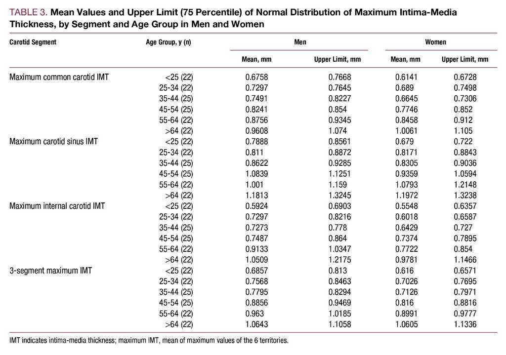
Ziel der Arbeit ist die Darstellung vaskulitisch bedingter Veränderungen der Arteria carotis interna infolge einer Riesenzellarteriitis (Arteriitis temporalis) Bei zwei von neun zwischen 1996 und 1998 konsekutiv, behandelten Patienten mit histologisch gesicherter Arteriitis temporalis fanden sich angiographisch für eine Vaskůlitis typische Veränderungen („vascular narrowings”) in. OBJECTIVE To reconcile various internal carotid artery (ICA) nomenclatures for transcranial and endoscopicendonasal perspectives, we reexamined the transition between lacerum (C3) and cavernous (C4) segments using a C1C7 segments schema In this cadaveric study, we obtained a 360°circumferential view integrating histological. Abstract Carotid intimamedia thickness (CIMT) has been shown to predict cardiovascular (CV) risk in multiple large studies Careful evaluation of CIMT studies reveals discrepancies in the comprehensiveness with which CIMT is assessedthe number of carotid segments evaluated (common carotid artery CCA, internal carotid artery ICA, or the carotid bulb), the type of measurements made (mean or maximum of single measurements, mean of the mean, or mean of the maximum for multiple.
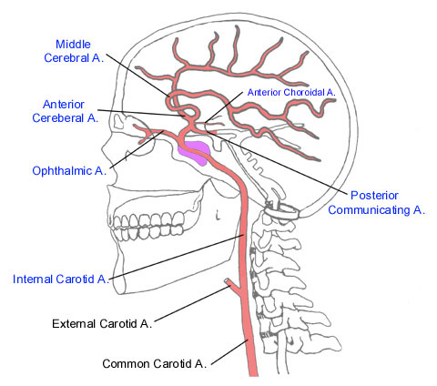
Arteria cerebri posterior — TA posterior cerebral artery divided into four segments the precommunicating part (pars precommunicalis), the postcommunicating part (pars postcommunicalis), the lateral occipital artery, and the medial occipital artery;. The internal carotid artery (C1 segment) enters the skull base through the carotid canal, where it begins a series of 90° turns which lead it to eventually terminate as the middle and anterior cerebral arteries. Internal carotid artery is one of the two terminal branches of common carotid artery It supplies structures present in the cranial cavity and orbit Its branches anastomose with the branches of external carotid artery in the scalp and face and middle ear Origin It begins at the upper border of the lamina of thyroid cartilage (level of disc between C3 and c4 vertebra).
But in the adult, the two vessels are of nearly equal size. Internal carotid artery is one of the two terminal branches of common carotid artery It supplies structures present in the cranial cavity and orbit Its branches anastomose with the branches of external carotid artery in the scalp and face and middle ear Origin It begins at the upper border of the lamina of thyroid cartilage (level of disc between C3 and c4 vertebra). Bilateral internal carotid artery agenesis is a rare lesion, with only 18 cases previously reported Blood supply to the anterior cerebral circulation is most commonly through enlarged basilar and.
The internal carotid artery (ICA) is a terminal branch of the common carotid artery Gross anatomy Origin It arises most frequently between C3 and C5 vertebral level, where the common carotid bifurcates to form the internal carotid and the external carotid artery (ECA) Variations in origin Although the majority arise between C3 and C5 vertebral level, a wide variation exists. The middle cerebral artery (MCA) is one of the three major paired arteries that supply blood to the brainThe MCA arises from the internal carotid artery (ICA) as the larger of the two main terminal branches (the other being the anterior cerebral artery), coursing laterally into the lateral sulcus where it branches and provides many branches that supply the cerebral cortex. Bilateral internal carotid artery agenesis is a rare lesion, with only 18 cases previously reported Blood supply to the anterior cerebral circulation is most commonly through enlarged basilar and.
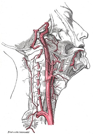
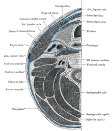
P1–P4 The P1 segment is the precommunicating part (pars precommunicalis), which begins at the bifurcation of the basilar artery and runs to the junction with the posterior communicating artery;. La arteria carótida interna es una rama terminal de la arteria carótida comúnNace aproximadamente al nivel de la tercera vértebra cervical, o en el borde superior del cartílago tiroides, cuando la carótida común se bifurca en esta arteria y la más superficial arteria carótida externa Desde su origen en el borde superior del cartílago tiroides (C4, o cuarta vértebra cervical), la. This part of the artery is known as the carotid sinus or the carotid bulb The ascending portion of the cervical segment occurs distal to the bulb, when the vessel walls are again parallel The internal carotid runs perpendicularly upward in the carotid sheath, and enters the skull through the carotid canal.
Infobox Artery Name = Internal carotid artery Latin = arteria carotis interna GraySubject = 146 GrayPage = 566 Width = 250 Caption = Arteries of the neck The. M3 is the opercular part, the segment emerging from the. OBJECTIVE To reconcile various internal carotid artery (ICA) nomenclatures for transcranial and endoscopicendonasal perspectives, we reexamined the transition between lacerum (C3) and cavernous (C4) segments using a C1C7 segments schema In this cadaveric study, we obtained a 360°circumferential view integrating histological.
A anterior cerebral artery (C7) The last two branches in the mnemonic are the terminal branches of the internal carotid artery Except for the terminal segment (C7) the oddnumbered segments usually have no branches, whereas the evennumbered segments (C2, C4, C6) each have two branches Calming Voices Make IntraOperative Surgery Pleasurable And Almost Memorable. In human anatomy, the internal carotid artery is a major artery of the head and neck that helps supply blood to the brain and is part of the circle of willis 1 Classification 2 Course 21 C1 Cervical segment 22 C2 Petrous segment 23 C3 Lacerum segment 24 C4 Cavernous part 25 C5 Clinoid. The Anterolateral Ganglionic Branches, a group of small arteries which arise at the commencement of the middle cerebral artery, are arranged in two sets one, the internal striate, passes upward through the inner segments of the lentiform nucleus, and supplies it, the caudate nucleus, and the internal capsule;.
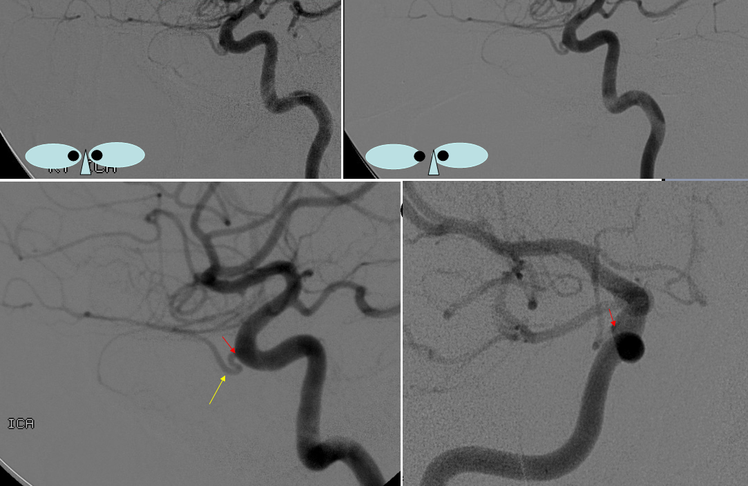
Branches Like every major artery in the human body, the internal carotid has several branches that are often asked about in anatomy exams They stem from several segments (C2, C4, C6, and C7), the only exceptions being the cervical (C1), lacerum (C3), and clinoid (C5) segments do not give rise to any branches. Variations of the course of the internal carotid artery in the parapharyngeal space and their frequency were studied in order to determine possible risks for acute haemorrhage during pharyngeal surgery and traumatic events, as well as their possible relevance to cerebrovascular disease The course of the internal carotid artery showed no curvature in 191 cases, but in 74 cases it had a medial. An obliquely resected segment of the internal carotid artery of appro priate length is excised so that the anastomotic lumen will be maximal The sectioned internal carotid artery is su tured endtoend to the common carotid artery.
Tętnica szyjna wewnętrzna (łac arteria carotis interna) – główne naczynie zaopatrujące przednią część mózgowia w krew tętniczą Biegnie od miejsca podziału tętnicy szyjnej wspólnej (34 kręg szyjny) do podstawy czaszki Przebieg Tętnica szyjna wewnętrzna biegnie przez szyję tranzytem nie oddając żadnych gałęzi. In anatomical terminology segments A1 (precommunicating part) and (postcommunicating part) are recognized In clinical terminology, the artery is described as comprising five. Tętnica szyjna wewnętrzna (łac arteria carotis interna) – główne naczynie zaopatrujące przednią część mózgowia w krew tętniczą Biegnie od miejsca podziału tętnicy szyjnej wspólnej (34 kręg szyjny) do podstawy czaszki Przebieg Tętnica szyjna wewnętrzna biegnie przez szyję tranzytem nie oddając żadnych gałęzi.
The primary role of the internal carotid artery is to deliver blood to the forebrain the front part of the brain that houses the cerebral hemispheres (which are involved higherlevel cognition, language, as well as visual processing), the thalamus (associated with visual, sensory, and auditory processing, sleep, and consciousness), and the hypothalamus (regulating metabolism and the release of hormones, among other functions). Internal carotid artery aka Arteria carotis interna in the latin terminology and part of arteries of the brain seen from the lateral and medial views of the brain This is an article about the segments, branches and clinical aspects of the internal carotid arteries Learn all about these important blood Arteria cerebri media. The four segments into which the middle cerebral artery is divided, M1â €“ M4 in clinical terminology The M1 segment is the sphenoid part (pars sphenoidalis), the horizontal portion of the vessel from the termination of the internal carotid artery to the point of branching at the limen insulae;.
Carotis Segments Images from MR Angiography (MRA) with Time of Flight (ToF) source sequence on the left and reconstructed Maximum Intensity Projection (MIP) showing stenoses in the Internal Carotid Artery (ICA) This patient has a stent in the distal carotid artery that beginns in petrous and ends in cavernous segment. An obliquely resected segment of the internal carotid artery of appro priate length is excised so that the anastomotic lumen will be maximal The sectioned internal carotid artery is su tured endtoend to the common carotid artery. Except for the terminal segment (C7) the odd numbered segments usually have no branches, whereas the even numbered segments (C2, C4, C6) each have two branches C1 cervical segment, none;.
Mnemonic C cervical segment P petrous segment L lacerum segment C cavernous segment C clinoid segment O ophthalmic segment C communicating segment. And C7, communicating This classification is practical, accounts for new anatomic information and clinical interests, and clarifies all segments of the ICA. The internal carotid artery (Fig 513) supplies the anterior part of the brain, the eye and its appendages, and sends branches to the forehead and nose Its size, in the adult, is equal to that of the external carotid, though, in the child, it is larger than that vessel It is remarkable for the number of curvatures that it presents in different parts of its course.
At approximately the level of the fourth cervical vertebra, the common carotid artery splits ("bifurcates" in literature) into an internal carotid artery (ICA) and an external carotid artery (ECA) While both branches travel upward, the internal carotid takes a deeper (more internal) path, eventually travelling up into the skull to supply the brain The external carotid artery travels more closely to the surface, and sends off numerous branches that supply the neck and face. 3 4 However, in clinical settings, the classification system of the internal carotid artery usually follows the 1996 recommendations by Bouthillier, 5 describing seven anatomical segments of the internal carotid artery, each with a corresponding alphanumeric identifier—C1 cervical, C2 petrous, C3 lacerum, C4 cavernous, C5 clinoid, C6 ophthalmic, and C7 communicating The Bouthillier nomenclature remains in widespread use by neurosurgeons, neuroradiologists and neurologists. Tętnica szyjna wewnętrzna (łac arteria carotis interna) – główne naczynie zaopatrujące przednią część mózgowia w krew tętniczą Biegnie od miejsca podziału tętnicy szyjnej wspólnej (34 kręg szyjny) do podstawy czaszki Przebieg Tętnica szyjna wewnętrzna biegnie przez szyję tranzytem nie oddając żadnych gałęzi.
C3 lacerum segment, none;. Bouthillier et al described (in 1996) a seven segment internal carotid artery (ICA) classification system It remains the most widely used system for describing ICA segments cervical segment C1;. 7 segments of Internal Carotid Artery We have already discussed a mnemonic to remember the course of Internal Carotid Artery with the help of 2 horizontal “S” under the topic of Circle of Willis C1 – Cervical segment C2 – Petrous (horizontal) segment C3 – Lacerum segment C4 – Cavernous segment C5 – Clinoid segment.
The segments into which the anterior cerebral artery is divided;. Arteria carotis interna kan motta blodforsyning via en viktig kollateral blodforsyning til hjernen, den cerebrale arterielle sirkel, bedre kjent som Willis' arterielle sirkel Grener Følgende liste navngir forgreningene til arteria carotis interna, sortert etter segment C1 Grener fra cervicalsegmentet – ingen.

C1 Cervical Segment Begins At The Level Of The Common Carotid Artery Download Scientific Diagram
:background_color(FFFFFF):format(jpeg)/images/article/en/middle-cerebral-artery/Z3kPNo9AeYobOFFpQWPWQ_Ozcn2x81beEbLydTy6reIg_A._cerebri_media__01.png)
Middle Cerebral Artery Anatomy Branches Supply Kenhub
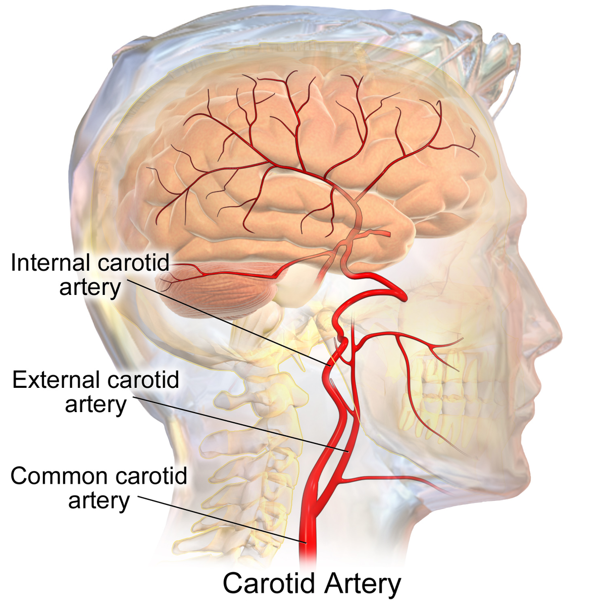
Internal Carotid Artery Wikipedia
Q Tbn And9gctllop1zf Fixomqtkvqtvoq3rcejf2iaeqvih Luncgjutzwhd Usqp Cau
1

Atypischer Abgang Pharyngookzipitaler Aste Aus Dem Zervikalen Segment Der Arteria Carotis Interna Springermedizin De

Carotid Siphon Geometry And Variants Of The Circle Of Willis In The Origin Of Carotid Aneurysms
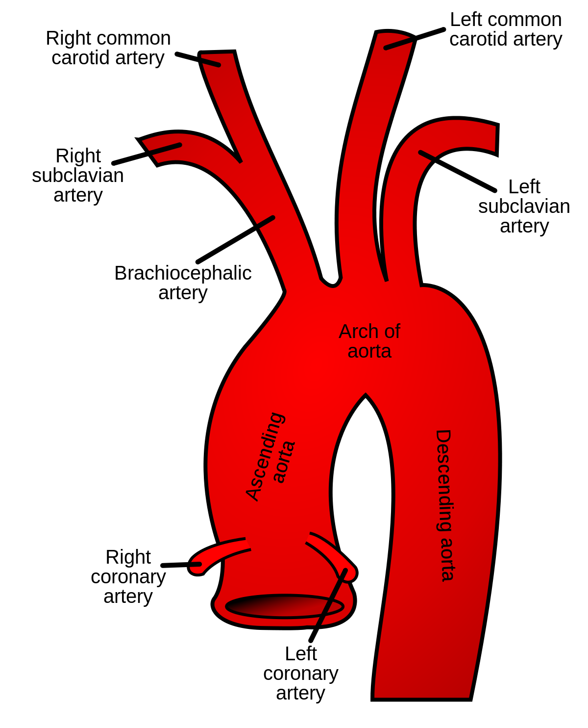
Common Carotid Artery Wikipedia

10 Best Internal Carotid Artery Ideas Carotid Artery Internal Carotid Artery Arteries

Illustration Of Cadaver Specimen 5 Depicts Single Origin Of The Download Scientific Diagram

Internal Carotid Artery Wikiwand
Internal Carotid Artery
Anat Lf1 Cuni Cz Souhrny Aofa6 Pdf
Internal Carotid Artery And Normal Variants

Carotid Siphon Geometry And Variants Of The Circle Of Willis In The Origin Of Carotid Aneurysms
Q Tbn And9gcrx0ssvbbfbngbstkt8tixjalcvkne4ltwgvwhtr4vkwl3lfhqx Usqp Cau

Pdf Carotid Artery Segmentation In Ultrasound Images And Measurement Of Intima Media Thickness Semantic Scholar
Internal Carotid Artery

External Carotid Artery Radiology Reference Article Radiopaedia Org

Segments Of Internal Carotid Artery
Anat Lf1 Cuni Cz Souhrny Aofa6 Pdf
:background_color(FFFFFF):format(jpeg)/images/article/en/internal-carotid-artery/6NDtFsuLG4yNmfWPFuBTQ_Internal_carotid_arteries.png)
Internal Carotid Artery Anatomy Segments And Branches Kenhub
:watermark(/images/watermark_5000_10percent.png,0,0,0):watermark(/images/logo_url.png,-10,-10,0):format(jpeg)/images/atlas_overview_image/2010/RPRXK47hgujCKnZyXSPlvw_3_english.jpg)
Internal Carotid Artery Anatomy Segments And Branches Kenhub

Pressure Drop In Tortuosity Kinking Of The Internal Carotid Artery Simulation And Clinical Investigation
Www Unispital Basel Ch Fileadmin Unispitalbaselch Bereiche Medizin Angiologie Fortbildungen Duplexsonografie19 De 2 1 Peters Duplex Normal 19 Folien Kompatibilit C3 tsmodus Pdf

Vascular Anatomy Neuroradiology Carotid Artery Arteries Anatomy Brain Anatomy
Anat Lf1 Cuni Cz Souhrny Aofa6 Pdf

Dangerous Extracranial Intracranial Anastomoses And Supply To The Cranial Nerves Vessels The Neurointerventionalist Needs To Know American Journal Of Neuroradiology

Hjarnans Blodkarl Neurologi

Collateral Variations In Circle Of Willis In Atherosclerotic Population Assessed By Means Of Transcranial Color Coded Duplex Ultrasonography Stroke

Internal Carotid Artery Wikipedia
Internal Carotid Artery Physiopedia

Cranial Arteries Of Moschiola Memmina A Left Lateral View B Download Scientific Diagram

Brain Blood Supply Position Structure Function Summary

Internal Carotid Artery Segments Branches Youtube

External Carotid Artery An Overview Sciencedirect Topics

Arteria Cerebri Interna Intracavernous Segments Neurosurgery
Internal Carotid Artery Segments And Branches Epomedicine
Http Www Clinicsinsurgery Com Pdfs Folder Cis V2 Id1551 Pdf
:watermark(/images/watermark_5000_10percent.png,0,0,0):watermark(/images/logo_url.png,-10,-10,0):format(jpeg)/images/atlas_overview_image/74/B81azdT3qAYziXueYQXqcA_arteries-of-brain-inferior-view_english.jpg)
Internal Carotid Artery Anatomy Segments And Branches Kenhub

Internal Carotid Artery And Its Aneurysms Neuroangio Org

Pin On Neuroradiology

Internal Carotid Artery And Its Aneurysms Neuroangio Org

Vernauwing Halsslagader Carotis Ziekte Info Neurologen Alrijne
Internal Carotid

Internal Carotid Artery And Normal Variants

Classification Of The Segments Of Circle Of Willis Cow Cow Segments Download Scientific Diagram

Pin On Neurology
External Carotid Artery Wikipedia

Simplified Arterial Tree Of Moschiola Memmina Illustrating Download Scientific Diagram

Detection Of Anomalous Cervical Internal Carotid Artery Branches By Colour Duplex Ultrasound European Journal Of Vascular And Endovascular Surgery
Www Journal Imab Bg Org Issues 17 Issue3 Jofimab 17 23 3p1657 1666 Pdf
Semmelweis Hu Anatomia Files 19 09 Blutgehirn Pdf
Circle Of Willis The Lecturio Medical Online Library
Internal Carotid Artery

Raeder S Syndrome After Embolization Of A Giant Intracavernous Carotid Artery Aneurysm Pathophysiological Considerations

A Carotis Interna Anatomie Eref Thieme
Http Www Clinicsinsurgery Com Pdfs Folder Cis V2 Id1551 Pdf
Internal Carotid Artery
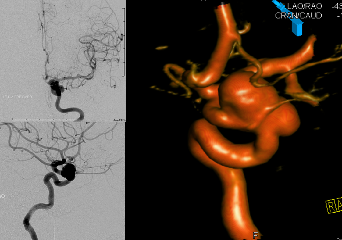
Internal Carotid Artery And Its Aneurysms Neuroangio Org
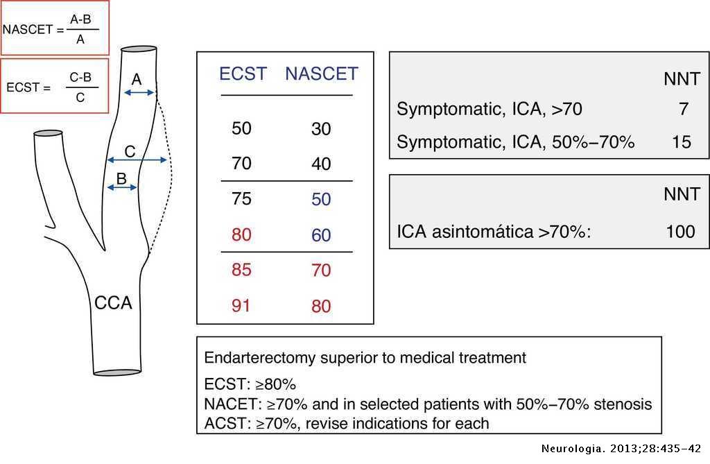
Ultrasound Measurement Of Carotid Stenosis Recommendations From The Spanish Society Of Neurosonology Neurologia English Edition
Arteria Cerebri Anterior Wikipedia
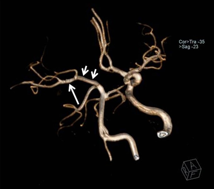
Einseitiges Fehlen Der Arteria Carotis Interna Springerlink

Internal Carotid Artery Anatomy Segments And Branches Kenhub
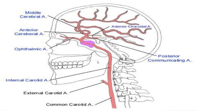
Segments Of Internal Carotid Artery
Anat Lf1 Cuni Cz Souhrny Alekzs1002b Pdf

Embolization Of Meningohypophyseal And Inferolateral Branches Of The Cavernous Internal Carotid Artery American Journal Of Neuroradiology
Internal Carotid Artery
Bouthiller Classification Of Internal Carotid Artery Anatomy Uw Emergency Radiology
Internal Carotid Artery
Internal Carotid Artery

Internal Carotid Artery Segments Radiologia Cabeca E Pescoco Cardiologia

Internal Carotid Artery Anatomy Circle Of Willis Youtube
Normal Vascular Anatomy Radiology Key
Http Www Ncbi Nlm Nih Gov Pmc Articles Pmc Pdf Joa 1973 0373 Pdf
Circle Of Willis Anatomy Overview Gross Anatomy Natural Variants
Internal Carotid Artery And Its Aneurysms Neuroangio Org

External Carotid Artery
Internal Carotid Artery Radiology Reference Article Radiopaedia Org

Raeder S Syndrome After Embolization Of A Giant Intracavernous Carotid Artery Aneurysm Pathophysiological Considerations

Anterior Circulation
Segments Of Internal Carotid Artery
Internal Carotid Artery
Anat Lf1 Cuni Cz Souhrny Alekzs1002b Pdf

Figure S 6 3d Reconstructie Van Een Nieuwe Pcoma Een Verbinding Download Scientific Diagram

Internal Carotid Artery Wikiwand
Internal Carotid Artery
:watermark(/images/watermark_5000_10percent.png,0,0,0):watermark(/images/logo_url.png,-10,-10,0):format(jpeg)/images/atlas_overview_image/1108/dVznzPzllzbyHTIf1orKQ_anatomy-hypophyseal-portal-system_english.jpg)
Internal Carotid Artery Anatomy Segments And Branches Kenhub

Internal Carotid Artery Wikipedia
Internal Carotid Artery Wikipedia
Carotid Intima Media Thickness In Subjects With No Cardiovascular Risk Factors Revista Espanola De Cardiologia

Pressure Drop In Tortuosity Kinking Of The Internal Carotid Artery Simulation And Clinical Investigation

Internal Carotid Artery Wikipedia
Anat Lf1 Cuni Cz Souhrny Aofa6 Pdf
Core Ac Uk Download Pdf Pdf
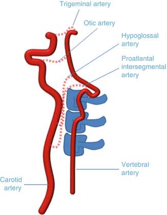
Imaging Of The Pathology Of The Vertebral Arteries Radiology Key

Internal Carotid Artery Radiology Reference Article Radiopaedia Org
2


