Meningeom Mrt T1
Klinik Und Poliklinik Fuer Neurochirurgie Meningeome
Www Thieme Connect De Products Ebooks Pdf 10 1055 B 0037 Pdf

Meningeom Wikiwand
Clinoid Meningioma Radiology Case Radiopaedia Org
Petroclival Meningiomas Radiological Features Essential For Surgeons Ecancer
Meningioma With Extension Through Skull Vault Radiology Case Radiopaedia Org
On occasion, a densely calcified meningioma may demonstrate hypointensity on T1 and T2weighted images;.

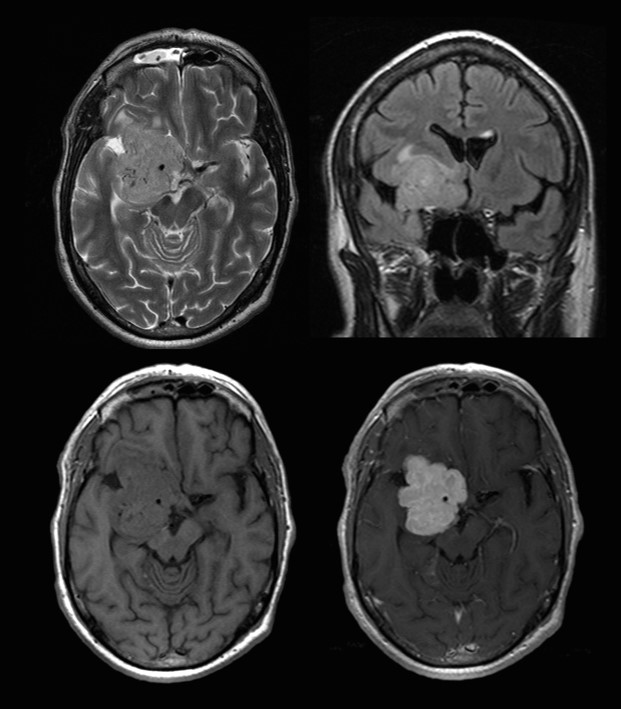
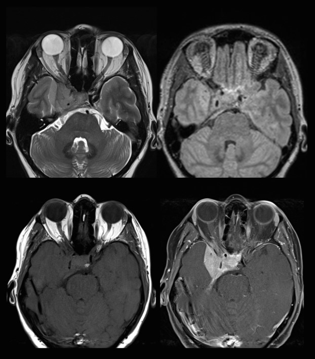
Meningeom mrt t1. The two basic types of MRI images are T1weighted and T2weighted images, often referred to as T1 and T2 images The timing of radiofrequency pulse sequences used to make T1 images results in images which highlight fat tissue within the body. Typical meningiomas appear as duralbased masses isointense to grey matter on both T1 and T2 weighted imaging enhancing vividly on both MRI and CT Some of the variants as mentioned earlier can, however, vary dramatically in their imaging appearance. In der T1Wichtung stellt sich Wasser hypointens dar, in der T2Wichtung ist es hyperintens!.
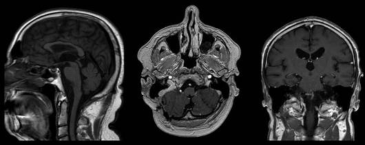
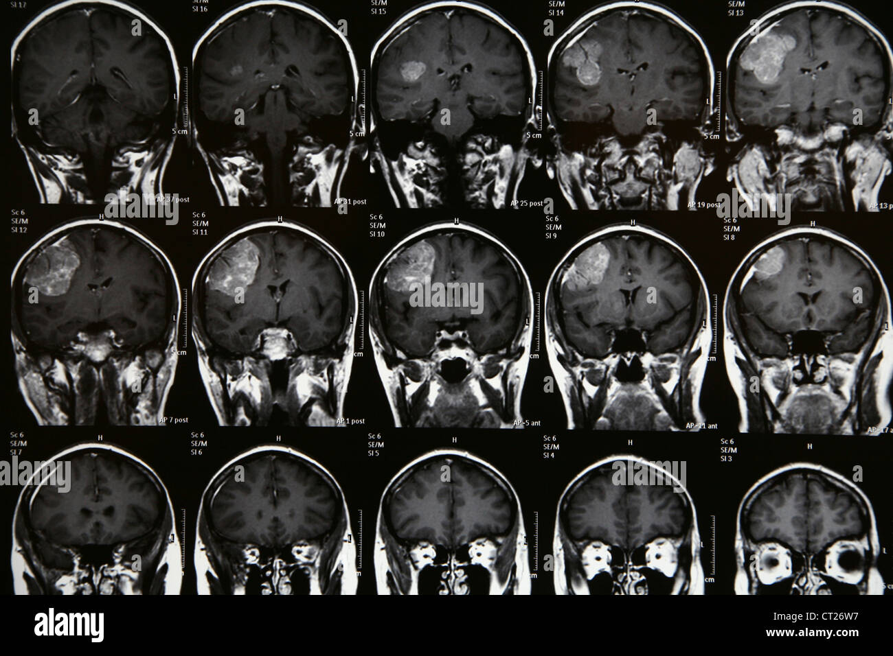
Coronal (A), transverse (B), and sagittal (C) gadoliniumenhanced T1weighted MRI shows bilateral olfactory meningiomas, and the falx dividing the tumor in 2 Arrow indicates tumor invasion of the sinuses D, Postoperative enhanced T1weighted MRI shows that the tumor was completely removed by means of craniotomy and a transfacial approach. Axial, sagittal, and coronal images reveal a round extraaxial lesion, with a broad base along the dura of the convexity, and a small associated dural tail Additional typical findings for the diagnosis include relative isointensity to brain on the T1weighted scan, slight hyperintensity on T2weighted scans, and homogeneous contrast enhancement. BACKGROUND AND PURPOSE The purpose of our study was to describe the MR imaging appearance of Warthin tumors multiple MR imaging techniques and to interpret the difference in appearance from that of malignant parotid tumors METHODS T1weighted, T2weighted, short inversion time inversion recovery, diffusionweighted, and contrastenhanced dynamic MR images of 19 Warthin tumors and 17.
38 yo m presents to ER for complaints of headaches for multiple months, worse in the morning Examination reveals multiple lipomas on arms, face, and legs MRI T1 post contrast reveals multiple meningiomas with asymptomatic hydrocephalus and cerebropontine angle mass Genetic work up in progress What surgical management and oncological management indicated?. On T1 and T2weighted MRIs, the tumors have variable signal intensity If a meningioma is suspected, obtaining an enhanced MRI is imperative Meningiomas enhance intensely and homogeneously after. A meningioma is a tumor that arises from the meninges — the membranes that surround your brain and spinal cord Although not technically a brain tumor, it is included in this category because it may compress or squeeze the adjacent brain, nerves and vessels Meningioma is the most common type of tumor that forms in the head.
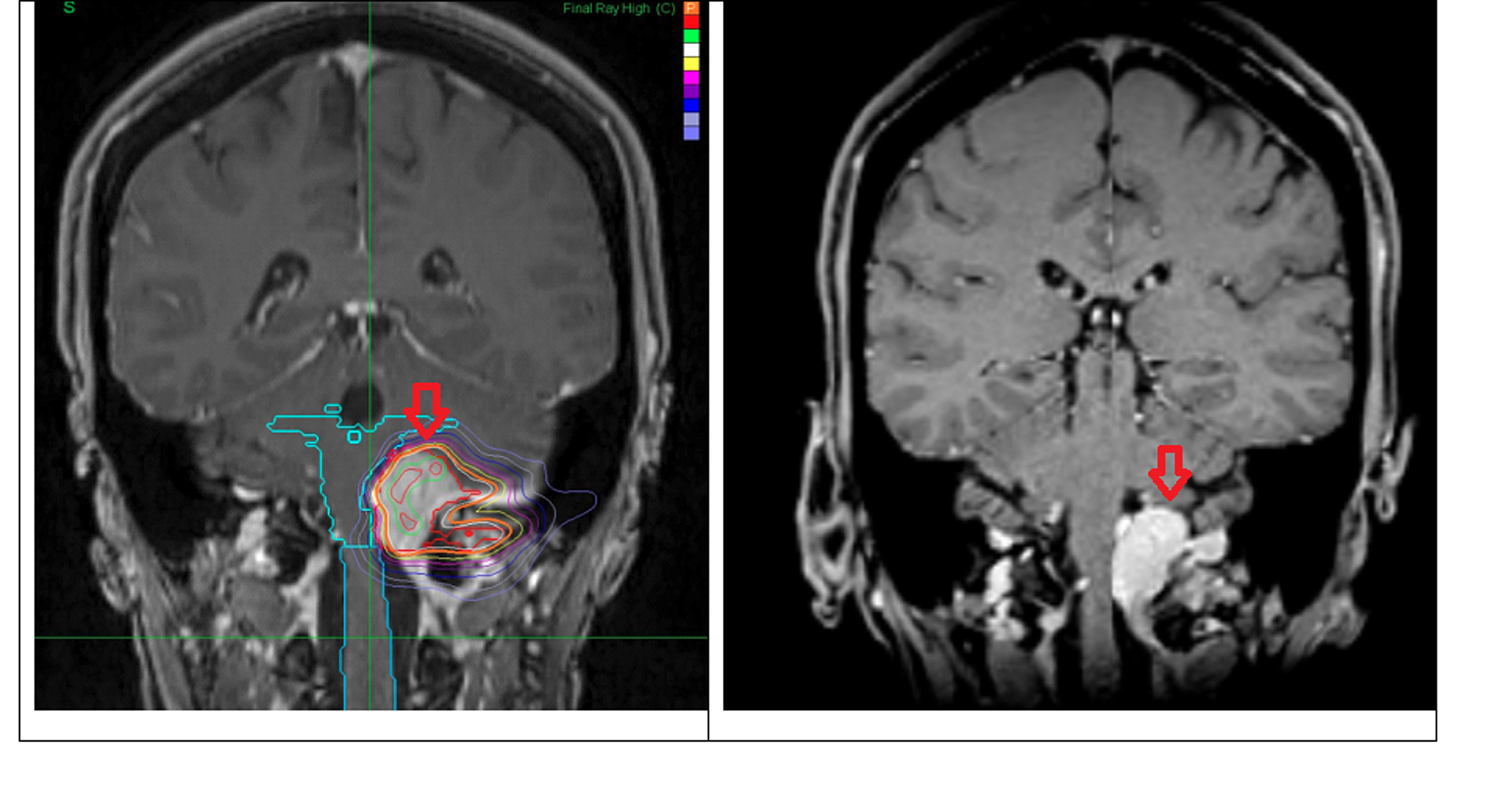
T1 reflects the length of time it takes for regrowth of Mz back toward its initial maximum value (Mo) Tissues with short T1's recover more quickly than those with long T1's Their Mz values are larger, producing a stronger signal and brighter spot on the MR image. Dr Amit M Shelat A Gamma knife unit uses 192 cobalt60 sources to emit gamma rays onto a preciselydefined target The precision of delivery of this radiation is less than 05 mm, oftentimes down to 025 mm. History 60 year old female with headaches and dizziness This is a case of a meningioma with all of the classic MRI findings, except for location Meningiomas arise from the arachnoid cap cells of the meninges They are supratentorial in 90% of the time, which is why this case isn't entirely a classical Meningiomas are benign.
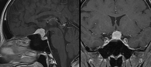
All patients were scanned with T1, T2weighted imaging (T1WI, T2WI), and contrastenhanced T1WI Results Convexity of brain was more likely to be involved, among 126 cases of meningioma, 45 (357%) tumors located at convexity of brain The size of tumor ranged from 14 to 99 cm Eightyone percent of tumors were round or oval in shape. Pituitary adenomas are primary tumors that occur in the pituitary gland and are one of the most common intracranial neoplasms Depending on their size they are broadly classified into pituitary microadenoma less than 10 mm in size;. Meningiomas are one of the most common extraaxial tumours of the central nervous system and accounts for 15 % of all intracranial neoplasms They are nonglial neoplasms that originate from the mesoderm or meninges Meningiomas are commonly found on the brain surface, either over the convexity or at the skull base.
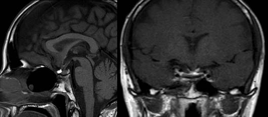
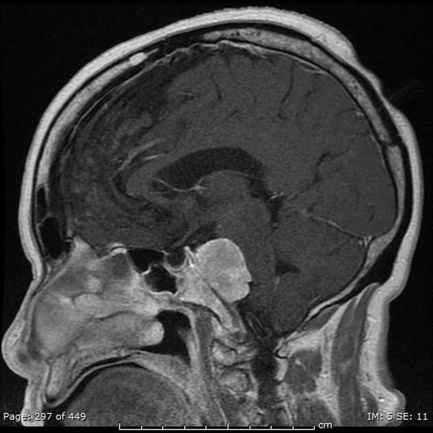
Image 2 sagittal T1_FL2D postcontrast (TR/TE 440/25 ms, scan time 2 min 26 sec, slice thickness 4 mm);. Although this distinction is largely arbitrary, it is commonly used and does highlight an important fact. The mass, isointense on T1 and T2weighted images (Fig 2), enhanced homogenously with intravenous gadolinium (Fig 3) The enhanced images also showed a dural attachment to the tumour and thickening of the dura mater at its origin, suggesting the diagnosis of meningioma Four vessel angiography showed neither abnormal vessels nor a tamour blush.
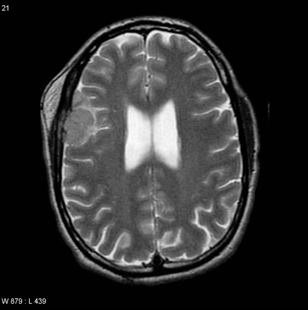
MR imaging and CT showed a welldemarcated intradiploic lesion with thickening of the skull extending from the frontal to the parietal calvarium with a low signal on T1weighted images, strong but. T1 enhanced T1weighted enhanced MRI image shows a hyperintense homogeneous round mass with an enhancing tail involving the dura. Inward displacement of the cortical.
H, Gadoliniumenhanced postoperative T1weighted MRI shows residual tumor, which was intentionally left to preserve patency of the SSS I, Pathology slide (hematoxylineosin, original magnification. Alternative Methoden Magnetresonanzspektroskopie Abkürzungen MRSpektroskopie, MRS;. Pituitary macroadenoma greater than 10 mm in size;.
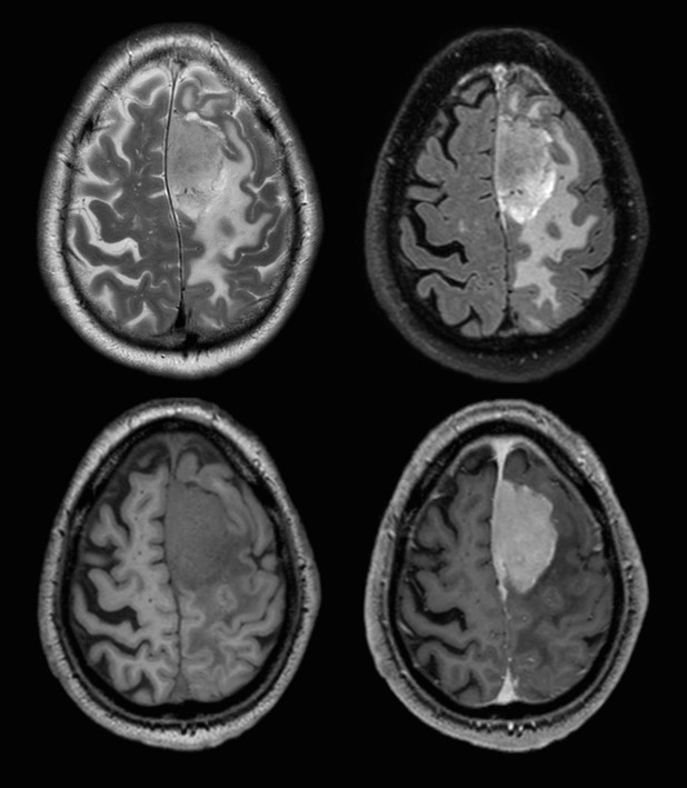
The MRI sequences demonstrate a wellcircumscribed broadbased left frontal extraaxial mass (4 x 35 x 25 cm) with adjacent hyperostosis of the inner table of the skull It shows an isosignal to the cortical grey matter on T1, T2, and FLAIR with areas of calcification of low signal on GE and mild surrounding vasogenic edema. Quattrocchi and his colleagues found this analysis conducted on a cohort of consecutive patients with meningioma and with no systemic interval therapy confirms that T1 signal hyperintensity of the dentate nucleus may be seen on unenhanced T1 images of patients undergoing multiple MRI scans with intravenous administration of gadodiamide. Coil dS HEAD Indskrivning MR CEREBRUM Lejring/centrering Pt ligger på ryggen Centrer på glabella Kontrast gives på lejet inden undersøgelsens start.
This page was last edited on 16 June 19, at 0809 Files are available under licenses specified on their description page All structured data from the file and property namespaces is available under the Creative Commons CC0 License;. Meningioma In the frontoparietal convexital area on T2weighted (a) and T1weighted MRI after intravenous CA administration (b, c), a flat spaceoccupying lesion is detected, intensely accumulating the contrast agent, infiltrating the adjacent bone, meninges and soft tissues of the frontoparietal region The tumor that has Vtype spread. 1 Definition Als T1Gewichtung bezeichnet man eine Kontrastdarstellung von MRT Bildern, bei der die Repetitionszeit (TR) und die Echozeit (TE) so gewählt werden, dass die untersuchten Gewebe vor allem durch ihre T1Relaxationszeit, und weniger ihre T2Relaxationszeit differenziert werden siehe auch T2Gewichtung 2 Physikalische Grundlagen.
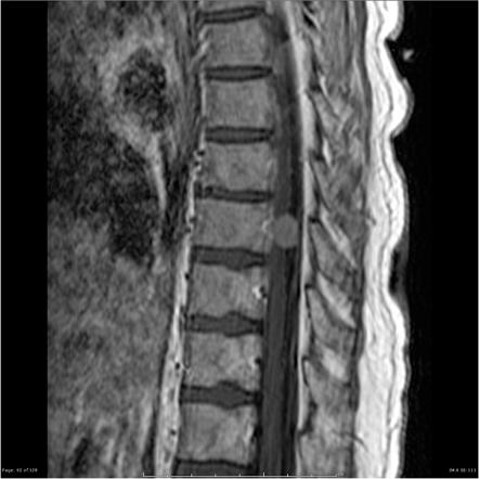
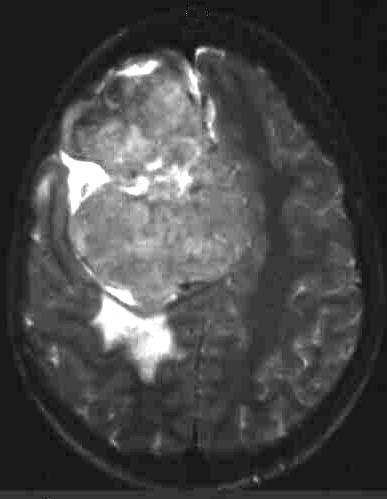
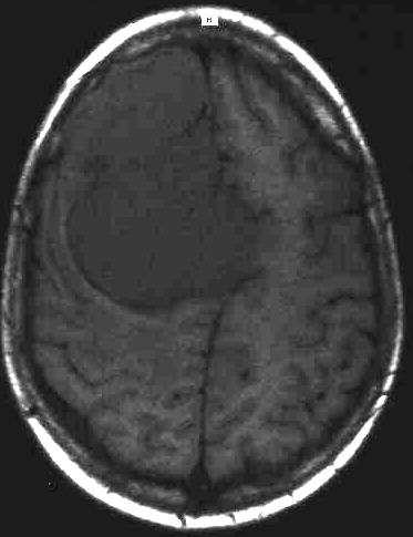
MRILibrary Hirnschädel Meningeom Indem Sie das Fenster schliessen (rechts oben anklicken) können Sie zur MRILibrary Auswahl zurückkehren T1gewichtetes MRT, T1gewichtetes MRT nach iv KM, T2gewichtetes MRT Transversale Schnittebene durch das Gehirn hochfrontal. Intraosseous meningioma, also referred to as primary intraosseous meningioma, is a rare subtype of meningioma that accounts for less than 1% of all osseous tumors They are the most common type of primary extradural meningiomas 6 Terminology I. Occasionally, densely calcified meningiomas are hypointense on T1 and T2 and show only minimal contrast enhancement Treatment and prognosis Spinal meningiomas are typically slow growing.
MRT 1 betrachten MRT 1 Schädelaufnahme (T1gewichtete Sequenz nach KM) Partielle Hemimegalencephalie links bei Tuberöser Hirnsklerose Große linke Hemisphäre mit großem linkem SV Verdickter, atypisch gyrierter Kortex und subkortikale Dysmyelinisierung (signalangehoben). Image 3 axial T1_FL2D postcontrast (TR/TE 250/25 ms, scan time 1 min 16 sec, slice thickness 4 mm) Imaging Findings. Rationale In the onecompartment model following iv administration the mean residence time (MRT) of a drug is always greater than its halflife (t(1/2)) However, following iv administration, drug plasma concentration (C) versus time (t) is best described by a twocompartment model or a two exponential equationC=Ae(alpha t)Be(beta t), where A and B are concentration unitcoefficients.
Eine entzündungsbedingte Knochenveränderung (zB Osteomyelitis, BrodieAbszess etc) zeigt im MRT das typische Signalmuster in der T1Wichtung dunkel, in der T2Wichtung hell, in der T1Wichtung nach der Gabe von Kontrastmittel hell Typisch ist auch der destruierende Charakter der Läsion, der Rand der Läsion ist diffus und unscharf. The lateral ventricle is markedly displaced (white arrow). Image 1 sagittal T1_FL2D precontrast (TR/TE 440/25 ms, scan time 2 min 26 sec, slice thickness 4 mm);.
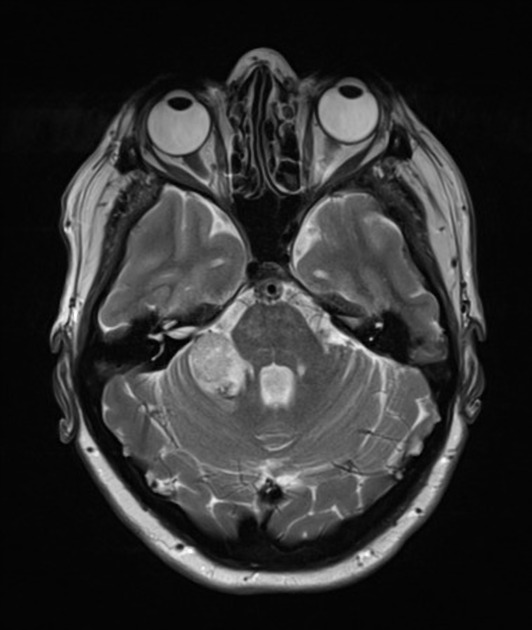
On March 11, 15, a retrospective study on 46 patients with a meningioma who had routinely undergone followup enhancedMRI scans with gadodiamide was published online in Investigative Radiology The authors report a significant increase in T1 hyperintensity of the dentate nuclei of the cerebellum on unenhanced scans was observed between the first and last MRI. Conventional MR imaging sequences are usually standard in many centers Multiplanar T1 and T2weighted images (WI), and postcontrast T1WI with 5 mm section thickness usually provide sufficient information to make a definitive radiologic diagnosis and a preoperative morphologic evaluation. On nonenhanced T1weighted images, most meningiomas have no signal intensity difference compared with cortical gray matter Fibromatous meningiomas may be more hypointense than the cerebral cortex T1weighted images may be used to asses for necrosis, hemorrhagic products, and cysts On T2weighted images, signal is variable.
Pearls A meningioma with intradural and extradural components occasionally mimics a nerve sheath tumor and a nerve sheath tumor with a predominant intradural component may mimic a meningioma;. The MRT station is situated at the basement of Terminal 2 You cannot help but find the way to Terminal 2 in order to get on board, no matter what terminal, 1, 2 or 3, you are arrived at Make it simple this way Know where you are (Terminal 1, 2 or 3) Follow the signs “Train to city” to get to the MRT station nestled at Terminal 2. T1 mapping refers to pixelwise illustrations of absolute T1 relaxation times on a map T1 mapping circumvents the influence of windowing and nulling (as in LGE) and allows direct T1 quantification As such, T1 mapping has the potential to detect diffuse myocardial structural alterations not assessable by other noninvasive means, including LGE.
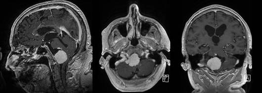
The following table shows T1 and T2 relaxation times for various tissues at 15 T For example A tissue with a long T1 and T2 (like water) is dark in the T1weighted image and bright in the T2weighted image A tissue with a short T1 and a long T2 is bright in the T1weighted image and gray in the T2weighted image. The risk of meningioma increases with age with a dramatic increase after 65 yearsChildren aged 014 are at the lowest risk African Americans have been observed to have higher rates of meningioma than other ethnic groups in the US Exposure to ionizing radiation, especially high doses, has been associated with a higher incidence of intracranial tumors, particularly meningiomas. A Die T1gewichteten MRTAufnahmen mit Kontrastmittel zeigen das große Meningeom mit Hirnstammkompression B Die postoperativen T1gewichtete MRTAufnahmen mit Kontrastmittel 1 Jahr postoperativ zeigen die komplette Tumorentfernung Der Hirnstamm liegt frei C und D Patientin 6 Monate postoperativ.
1 Definition Als T1Gewichtung bezeichnet man eine Kontrastdarstellung von MRTBildern, bei der die Repetitionszeit (TR) und die Echozeit (TE) so gewählt werden, dass die untersuchten Gewebe vor allem durch ihre T1Relaxationszeit, und weniger ihre T2Relaxationszeit differenziert werden siehe auch T2Gewichtung 2 Physikalische Grundlagen Bei jeder Bildakquisition wird die gewählte. The mass is isointense on T1weighted images but strongly enhances with contrast This is the typical appearance of a meningioma Meningiomas are common tumors that arise from the meninges and can occur within the spinal canal as well as intracranially They are typically benign histologically, and can be surgically resected if they are in an. A Die T1gewichteten MRTAufnahmen mit Kontrastmittel zeigen das große Meningeom mit Hirnstammkompression B Die postoperativen T1gewichtete MRTAufnahmen mit Kontrastmittel 1 Jahr postoperativ zeigen die komplette Tumorentfernung Der Hirnstamm liegt frei C und D Patientin 6 Monate postoperativ.
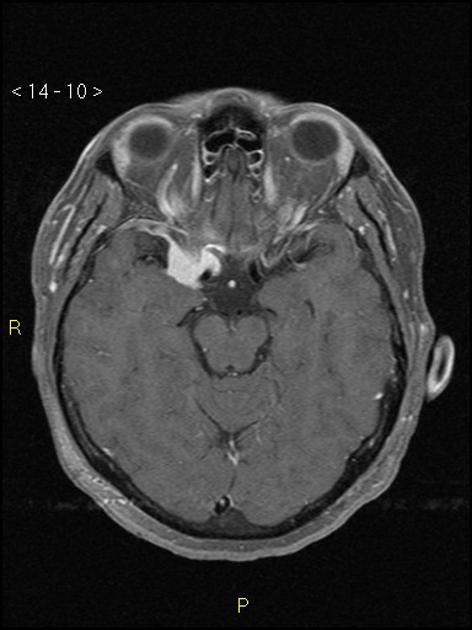
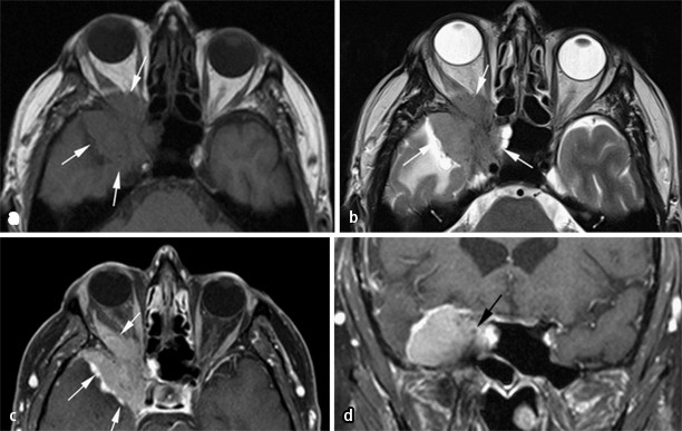
MRT 1 T1Wichtung koronar nach KMGabe vor Therapie deutliche Verbreiterung (ca 1cm) und pathologisches Kontrastmittelenhancement meningealer Strukturen parietal und hochparietal bds MRmorphologisch kann nicht sicher zwischen einem duralen oder leptomeningealen Befall unterschieden werden, jedoch deutet die glatte Kontur nach. Axial T1 An intensely high T2 signal extraaxial mass with vivid contrast enhancement and a dural tail (best seen on coronal) is seen in the left middle cranial fossa Case Discussion. Axial T1 and T2w MRI sections show an extra axial dura based solid signal intensity mass, overlying lateral sphenoid wing, homogenously iso intense to cortical grey matter on T1 and T2w images, intense homogenous enhancement on post contrast T1 with dural tailing Marked peri lesional Odema in adjacent compressed left frontal lobe parenchyma.
Magnetic resonance images of an olfactory and frontal lobe meningioma with a large eccentrically located cystic structure (A) Parasagittal postcontrast T1‐weighted (T1W) image showing a uniformly contrast enhancing meningioma with an associated cyst (black arrow) extending caudally;. Meningiomas may appear different on T1 and T2weighted sequence but with a few similarities MRI Head MRI may be helpful in the diagnosis of meningioma Findings on MRI suggestive of/diagnostic of meningioma include Lobular, extra axial masses with wellcircumscribed margins;. However, nerve sheath tumors are usually.
Long T1 signal presents obvious enhanced, and the range of enhancement is extended D (HE stain, magnification times × 10) The image exhibits the transition or mixture between meningothelial and fibroblastic meningiomas The tumor cells have a lobulated or fascicular arrangement with large numbers of swirling structures. A meningioma is a tumor that grows in the protective lining of the brain and spinal cord, called the meninges Most are benign, though in rare cases they can be cancerous (malignant). Nativ im MRT T1 → isointens, T2 → iso bis hyperintens Sonderform Meningeom en plaque Meningeom der mittleren Schädelgrube mit starker ossärer Komponente In Ausnahmefällen Probebiopsie mit Bestimmung der Histologie.
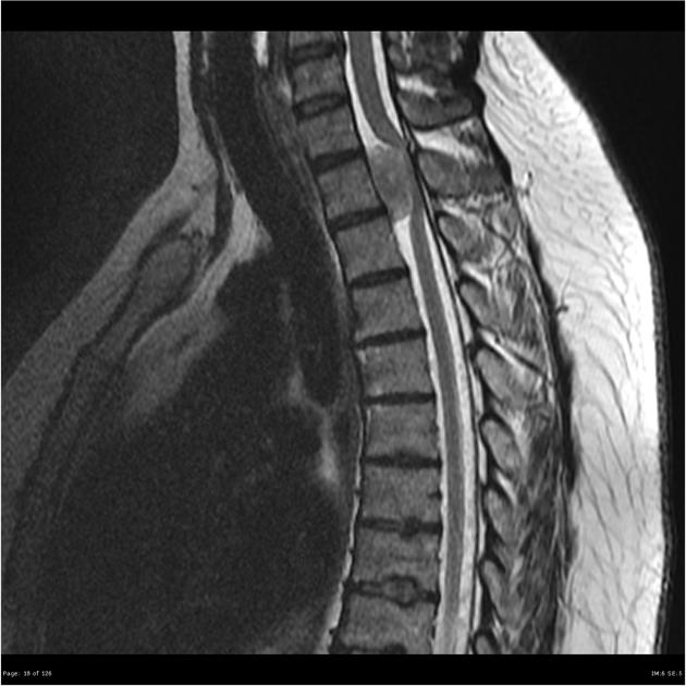
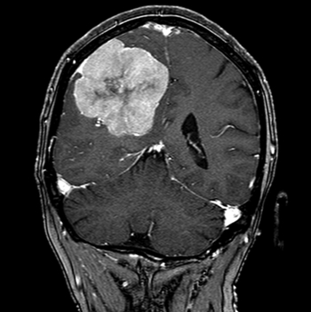
Auf dieser Abbildung sieht man ein Meningeom im T1gewichteten MRT nach Kontrastmittelgabe Ein 76jähriger Mann klagte über Übelkeit, Erbrechen, Synkopen, Sensibilitätsstörungen und eine Aphasie Bei dem daraufhin durchgeführten kranialen CT entdeckte man einen 6 cm großen Tumor im frontalen Kortex. Definition Verfahren, das den Spin von unterschiedlichen Protonen in einem definierten. Sagittal T1W MR image of the cervical spine reveals a large T1 isointense mass at the level of the foramen magnum (arrow) Even on this T1W image, at the superior portion of the mass there is a dural “tail” Sagittal T2W MR image in the same patient shows the mass is markedly T2 hypointense.
A comparison of the MRI specific acronyms for magnetic resonance imaging sequences, eg TSE, FSE, FLAIR, STIR, FLASH, and FISP, used by the manufacturers GE, Philips. Additional terms may apply. Methods We recruited 131 patients with pathological diagnosis of meningiomas All the patients had undergone MRI before surgery on a 30 T MRI scanner to obtain T1 fluid attenuated inversion recovery (T1 FLAIR) images, T2weighted images (T2WI) and T1 FLAIR with contrast enhancement (CET1 FLAIR) images covering the whole brain.
MRILibrary Hirnschädel Meningeom Indem Sie das Fenster schliessen (rechts oben anklicken) können Sie zur MRILibrary Auswahl zurückkehren T1gewichtetes MRT, T1gewichtetes MRT nach iv KM, T2gewichtetes MRT Transversale Schnittebene durch das Gehirn hochfrontal. T1 T2 postkontrastne Meningeóm angiografia 50 ročná žena, fajčiarka, hypertonička, diabetička Pred 2 rokmi začala mať ráno bolesti hlavy, ktoré spontánne ustúpili Asi pred rokom začala mať problémy so sluchom vľavo, pravidelne sa k hovoriacim otáčala pravou stranou. MRI was performed as an axial threedimensional gradientecho T1weighted sequence at 16 mm slice thickness without gap (3DMRI) Results were compared to the reports of diagnostic findings Mean tumor size of all 59 meningiomas was 139 ml (08–629 ml) before treatment.
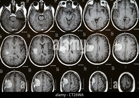
An axial, T1weighted MRI scan (right) reveals a homogenously enhancing, duralbased lesion, the classic appearance of a meningioma Images courtesy of Anand Rughani, MD While many meningiomas are. Conventional MR imaging sequences are usually standard in many centers Multiplanar T1 and T2weighted images (WI), and postcontrast T1WI with 5 mm section thickness usually provide sufficient information to make a definitive radiologic diagnosis and a preoperative morphologic evaluation. Coronal T1 shows intermediate to high signal in the plexus cyst Note also low signal in the right plexus on T2 (old hemorrhage) and high signal on FLAIR in the left plexus (acute hemorrhage) Choroid Plexus Cysts are very common normal finding Those often show restricted diffusion However in our case of patient with hypertension this finding.
In this work, the brain lesions that cause spontaneously hyperintense T1 signal on MRI were studied under seven categories The first category includes lesions with hemorrhagic components, such as infarct, encephalitis, intraparenchymal hematoma, cortical contusion, diffuse axonal injury, subarachnoid hemorrhage, subdural and epidural hematoma, intraventricular hemorrhage, vascular. Merksprüche „T1 und T2 ist wie SchwarzWeißSehen von Flüssigkeiten“ und „ H 2 O ist in T 2 hyperintens (h ell)“!. The mass is isointense on T1weighted images but strongly enhances with contrast This is the typical appearance of a meningioma Meningiomas are common tumors that arise from the meninges and can occur within the spinal canal as well as intracranially They are typically benign histologically, and can be surgically resected if they are in an.
Http Www Uksh De Uksh Media Dateien Kliniken Institute Radiologiezentrum Neuroradiologie Hl Dokumente Intrakranielle Blutungen Pdf

Meningeom Des Gehirns Eref Thieme
Q Tbn And9gcrq xppbksqcinlketi18u5jnknkpc1hbkyhde6v4rjz2ieg6 Usqp Cau
Spinal Meningioma Radiology Reference Article Radiopaedia Org
Bilder Buecher De Zusatz 772 7784 Lese 1 Pdf
Cureus Unresectable Foramen Magnum Meningioma Treated With Cyberknife Robotic Stereotactic Radiosurgery

Meningeom Des Gehirns Eref Thieme

Meningeom Des Gehirns Eref Thieme
Www Uniklinikum Leipzig De Einrichtungen Neurochirurgie Freigegebene dokumente Ausbildungsinhalte Neurochirurgie Uniklinikum Leipzig Pdf
Www Thieme Connect De Products Ebooks Pdf 10 1055 B 0037 Pdf
Extraaxiale Hirntumoren Springerlink
Http Www Unimedizin Mainz De Fileadmin Kliniken Neurochirurgie Dokumente Lehre Kv Meningeom Ws 18 19 Pdf
Figure 2 Radiologische Diagnostik Von Meningeomen Springerlink
Schweizer Hirntumorstiftung Zurich Stiftung Fur Hirntumor Patienten Krebsbekampfung Meningeom Who Grad Iii

Radiologie Flashcards Quizlet
Klinik Und Poliklinik Fuer Neurochirurgie Meningeome

Fallsammlung Radiologie Neuroradiologie Berlin Mitte Dr Korves Smartphone Tauglich
Klinik Und Poliklinik Fuer Neurochirurgie Meningeome

Meningeom Des Gehirns Eref Thieme

Planum Sphenoidale Meningeom Ars Neurochirurgica

Figure 1 Radiologische Diagnostik Von Meningeomen Springerlink
Http Www Unimedizin Mainz De Fileadmin Kliniken Neurochirurgie Dokumente Lehre Kv Meningeom Ws 18 19 Pdf
Bilder Buecher De Zusatz 772 7784 Lese 1 Pdf

Mri Of Orbit Case 1 A Axial T1 Weighted B Axial T1 Weighted Download Scientific Diagram
Tentorium Cerebelli Meningioma Radiology Case Radiopaedia Org
Http Www Unimedizin Mainz De Fileadmin Kliniken Neurochirurgie Dokumente Lehre Kv Meningeom Ws 18 19 Pdf
Mri Library Meningeom

Meckel Cave Meningioma Radiology Case Radiopaedia Org
Meningeom Mrt Stockfotografie Alamy
Meningeom Mrt Stockfotografie Alamy

Hirntumore Was Taugen Mrt Bilder Und Radiologische Befunde Pdf Free Download

Meningeom Des Gehirns Eref Thieme
Meningioma Of The Clivus Radiology Case Radiopaedia Org
Schweizer Hirntumorstiftung Zurich Stiftung Fur Hirntumor Patienten Krebsbekampfung Meningeom Who Grad I

Coronal Gadolinium Dtpaenhanced T1 Weighted Mri Slices Of Srs Op4 Download Scientific Diagram
Link Springer Com Content Pdf 10 1007 2f978 3 7091 6314 6 5 Pdf

Neurorad 19 Automatische Mrt Basierte Meningeom Segmenti

Neurorad 19 Automatische Mrt Basierte Meningeom Segmenti

Uberweisung Untersuchung Kontrastmittel Labor Radiologie Neuroradiologie Berlin Mitte Dr Korves Smartphone Tauglich
Schweizer Hirntumorstiftung Zurich Stiftung Fur Hirntumor Patienten Krebsbekampfung Meningeom Who Grad Ii
Klinik Und Poliklinik Fuer Neurochirurgie Meningeome
Klinik Und Poliklinik Fuer Neurochirurgie Meningeome
Www Neurorad Gr Image Ashx I Pdf Fn 11 Pdf
Www Ukm De Fileadmin Ukminternet Daten Kliniken Radiologie Pdf Neuroradiologie Pdf

Therapie Uniklinik Tubingen Neurochirurgie
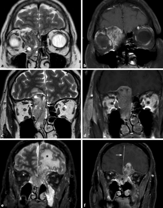
Bildgebung Der Nasennebenhohlen Und Der Frontobasis Springerlink
Http Www Unimedizin Mainz De Fileadmin Kliniken Neurochirurgie Dokumente Lehre Kv Meningeom Ws 18 19 Pdf
Www Neurorad Gr Image Ashx I Pdf Fn 17 Pdf
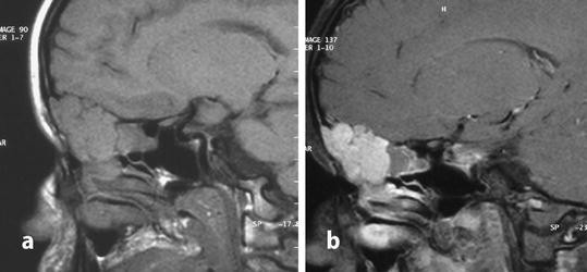
Tumoren Der Schadelbasis Springerlink

Meningioma Mri Wikidoc
Http Www Unimedizin Mainz De Fileadmin Kliniken Neurochirurgie Dokumente Lehre Kv Meningeom Ws 18 19 Pdf
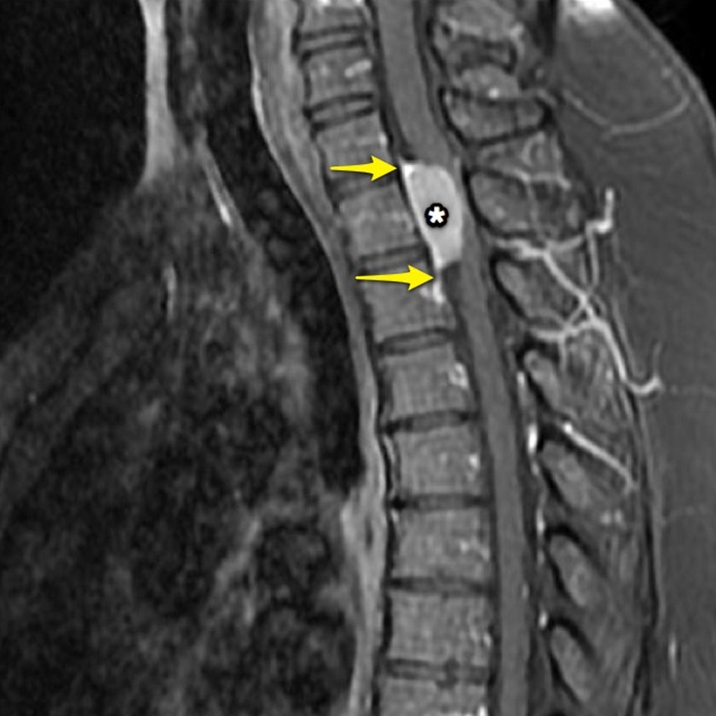
Spinal Meningioma Radiology Case Radiopaedia Org
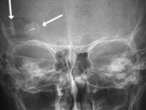
Klinik Und Poliklinik Fuer Neurochirurgie Meningeome

Mri Library Hyperperfusion Eines Meningeoms
Nanopdf Com Download Skript Tumore Pdf
Www Neurorad Gr Image Ashx I Pdf Fn 17 Pdf

The Diagnostic Value Of Diffusion Weighted Imaging In Patients With Meningioma Sciencedirect
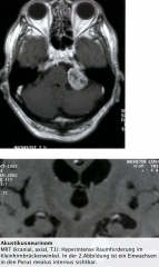
Staats Neurologie Hirntumore Foreign Language Flashcards Cram Com

Uberweisung Untersuchung Kontrastmittel Labor Radiologie Neuroradiologie Berlin Mitte Dr Korves Smartphone Tauglich
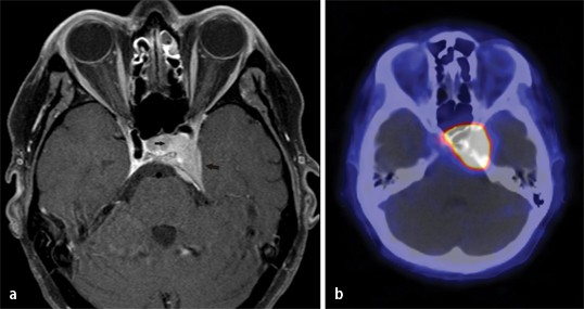
Figure 6 Radiologische Diagnostik Von Meningeomen Springerlink
Www Thieme Connect De Products Ebooks Pdf 10 1055 B 0034 Pdf

Meningioma Radiology Key
Mri Library Meningeom
Q Tbn And9gcr4hcy5t Sj0e 7cwbg4y4jy0y35v Gjj77osonmq1ubhukgzq Usqp Cau
D Nb Info 34

Meningioma En Plaque Radiology Case Radiopaedia Org
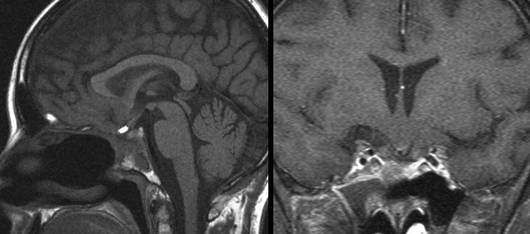
Klinik Und Poliklinik Fuer Neurochirurgie Meningeome

Extraaxiale Hirntumoren Springerlink
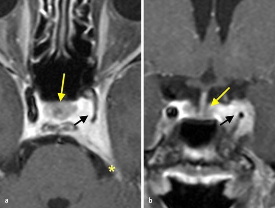
Tumoren Der Sellaregion Springerlink

Meningeom Des Gehirns Eref Thieme

File Falxmeningeom Mrt T1 Mit Kontrastmittel Jpg Wikimedia Commons
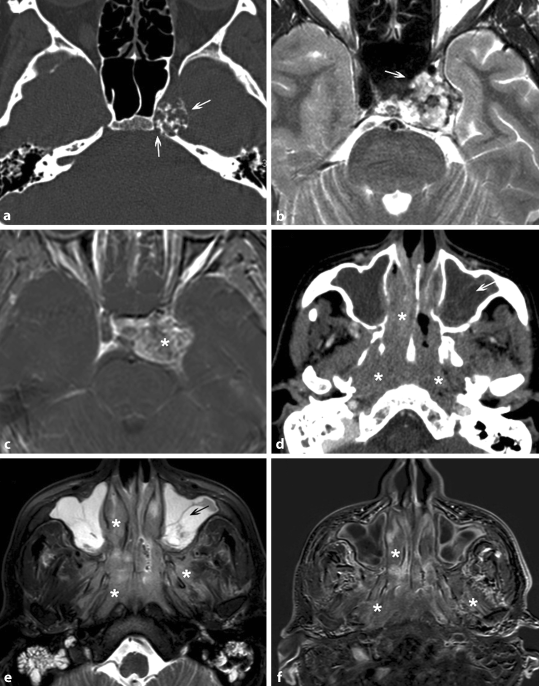
Bildgebung Der Nasennebenhohlen Und Der Frontobasis Springerlink
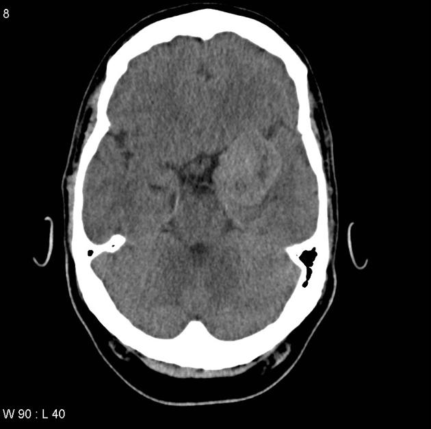
Meningioma Sphenoid Wing Typical Radiology Case Radiopaedia Org
Bilder Buecher De Zusatz 772 7784 Lese 1 Pdf

Grundlagen Und Unterscheidungsmerkmale Zu Wichtigen Mrt Sequenzen Youtube

Pdf Spinale Tumoren
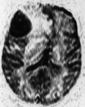
Mri Library Hyperperfusion Eines Meningeoms
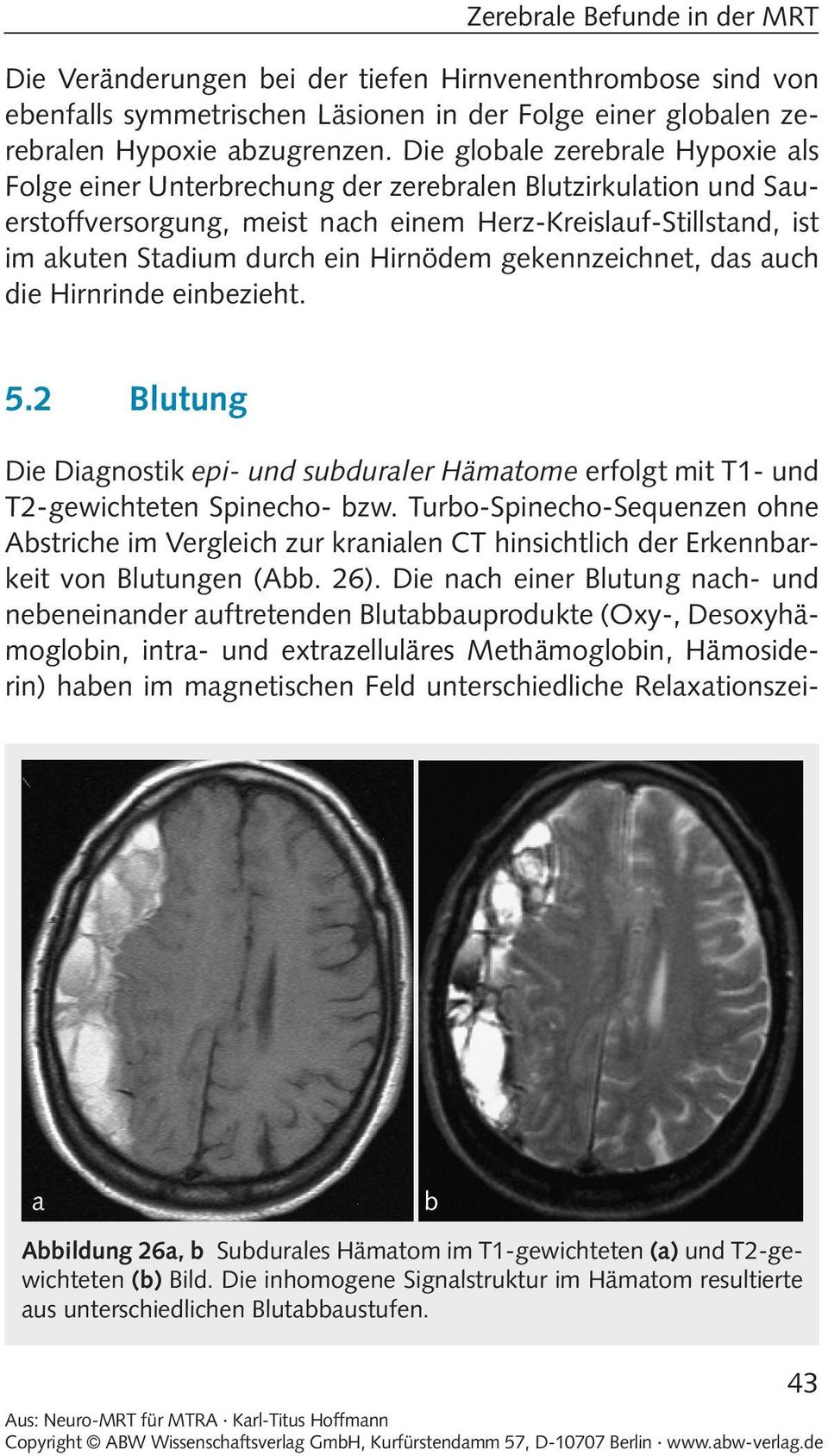
Blutung Zerebrale Befunde In Der Mrt Pdf Free Download
Nanopdf Com Download Skript Tumore Pdf

Bildgebende Diagnostik Des Zns Via Medici Leichter Lernen Mehr Verstehen
Nanopdf Com Download Skript Tumore Pdf
Q Tbn And9gcs4ppmgnb3zpavm1yulhrk0ub6heciy 9 W gb0qsc Iwhoid Usqp Cau

Meningioma Radiology Key
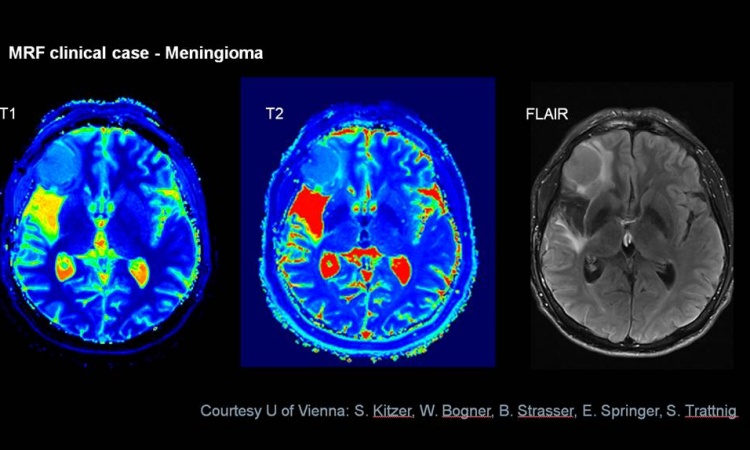
The Impact Of A Radiological Transformation
Www Thieme Connect De Products Ebooks Pdf 10 1055 B 0037 Pdf
Www Thieme Connect De Products Ebooks Pdf 10 1055 B 0036 Pdf

Mri Of Orbit Case 1 A Axial T1 Weighted B Axial T1 Weighted Download Scientific Diagram
Www Neurorad Gr Image Ashx I Pdf Fn 11 Pdf
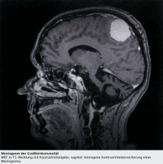
Staats Neurologie Hirntumore Foreign Language Flashcards Cram Com

Uberweisung Untersuchung Kontrastmittel Labor Radiologie Neuroradiologie Berlin Mitte Dr Korves Smartphone Tauglich
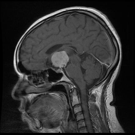
Suprasellar Meningioma Radiology Case Radiopaedia Org

Meningeom Der Wirbelsaule Eref Thieme
Q Tbn And9gcrlf7oefeyk9cspp2twbp Qjkplprqneb6cfs8v7io Usqp Cau

Meningioma Bright On T2 Radiology Case Radiopaedia Org
Http Www Unimedizin Mainz De Fileadmin Kliniken Neurochirurgie Dokumente Lehre Kv Meningeom Ws 18 19 Pdf

File Meningeom Im Spinalkanal Mrt Jpg Wikimedia Commons
Spinal Meningioma Radiology Case Radiopaedia Org
Neuroradiologische Diagnostik Und Therapie Radiologie Lubeck Sana Kliniken Ag


