Soft Agar Colony Formation Assay

Inhibitory Effect Of Hybrid Liposomes On The Growth Of Liver Cancer Stem Cells Sciencedirect

Cytoselect 96 Well In Vitro Tumor Sensitivity Assay Soft Agar Colony Formation Assay Kit

Cytoselect 96 Well In Vitro Tumor Sensitivity Assay Soft Agar Colony Formation Assay Kit

The Prostate Cancer Risk Variant Rs Regulates Multiple Gene Expression Through Extreme Long Range Chromatin Interaction To Control Tumor Progression Science Advances
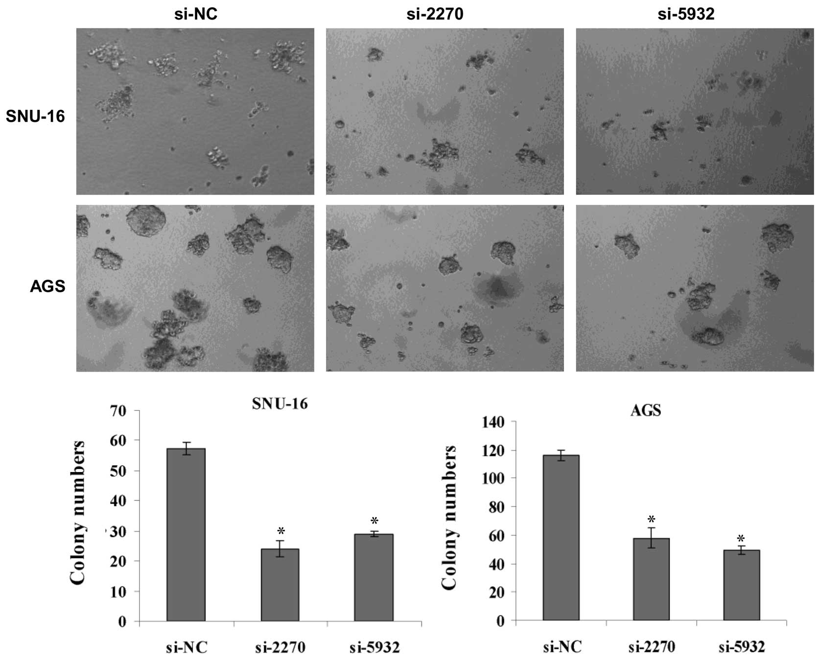
Knockdown Of Hmex 3a By Small Rna Interference Suppresses Cell Proliferation And Migration In Human Gastric Cancer Cells
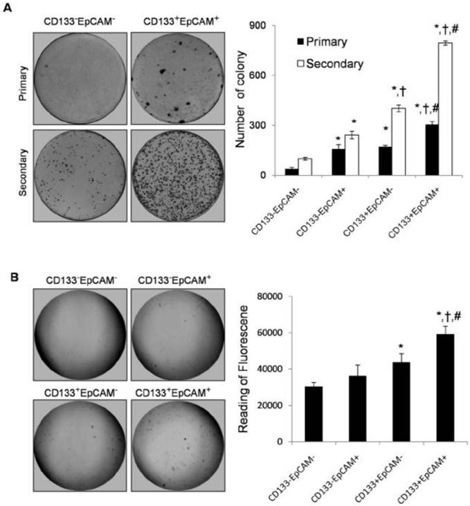
Cd133 Epcam Phenotype Possesses More Characteristics Of Tumor Initiating Cells In Hepatocellular Carcinoma Huh7 Cells
The Soft Agar Assay for Colony Formation is an anchorage independent growth assay in soft agar, which is considered the most stringent assay for detecting malignant transformation of cells For this assay, cells (pretreated with carcinogens or carcinogen inhibitors) are cultured with appropriate controls in soft agar medium for 2128 days.

Soft agar colony formation assay. Soft agar assay for colony formation September 3, 15 / 0 Comments / in Molecular Biology , Protocols / by admin Note All volumes are calculated to cater for four plates per point. The soft agar colony formation assay has traditionally been used to monitor ancorageindependent growth, employing 34 weeks of cell growth followed by manuel cell counting We have advanced the soft agar assay to eliminate tedious manual cell counting, allow highthroughput drug screening, and enable recovery of transformed cells for downstream. In these cases, see Protocol Measuring Survival of Hematopoietic Cancer Cells with the ColonyForming Assay in Soft Agar (Crowley and Waterhouse 15a).
Soft Agar Colony Formation The soft agar colony formation assay was performed according to our previous publication Briefly, a heterogeneous mixture of 5 × 10 3 cells in 03 mL of 04% agar medium was layered on top of 05 mL of 08% agar medium in a 24well plate PL54R, PL54S, and PL54Rac were each added to the top agar at the desired. A clonogenic assay is a cell biology technique for studying the effectiveness of specific agents on the survival and proliferation of cells It is frequently used in cancer research laboratories to determine the effect of drugs or radiation on proliferating tumor cells as well as for titration of Cellkilling Particles (CKPs) in virus stocks It was first developed by TT Puck and Philip I. The soft agar colony formation assay is a wellestablished method for characterizing this capability in vitro and is considered to be one of the most stringent tests for malignant transformation in cells This assay also allows for semiquantitative evaluation of this capability in response to various treatment conditions.
For each 35 mm dish, mix 750 µL of cells in 2x medium 750 µL of 06% agar to give you 15 mL of 03% agar in 1x medium containing 5000 cells, then immediately plate on top of base agar Assays are usually done in triplicate, so account for 3 wells/condition in calculations;. The soft agar colony formation assay is a method used to confirm cellular anchorageindependent growth in vitro The goal of this protocol is to illustrate a stringent method for the detection of the tumorigenic potential of transformed cells and the tumor suppressive effects of proteins on transformed cells. The soft agar colony formation assay is a method used to confirm cellular anchorageindependent growth in vitro The goal of this protocol is to illustrate a stringent method for the detection of the tumorigenic potential of transformed cells and the tumor suppressive effects of proteins on transformed cells.
Soft agar colony formation assay is a wellestablished method to evaluate cellular anchorageindependent growth for the detection of the tumorigenic potential of malignant cells (Roberts et al, 1985), which is developed from plate colony formation assay described by Puck et al in 1956 where cells were seeded on to a culture plate to assay the ability of cells to form colonies (Puck et al, 1956). Soft Agar Colony Formation The soft agar colony formation assay was performed according to our previous publication Briefly, a heterogeneous mixture of 5 × 10 3 cells in 03 mL of 04% agar medium was layered on top of 05 mL of 08% agar medium in a 24well plate PL54R, PL54S, and PL54Rac were each added to the top agar at the desired concentration. Given the inherent difficulties in investigating the mechanisms of tumor progression in vivo, cellbased assays such as the soft agar colony formation assay (hereafter called soft agar assay), which measures the ability of cells to proliferate in semisolid matrices, remain a hallmark of cancer research.
The soft agar colony formation assay is a wellestablished method for characterizing this capability in vitro and is considered to be one of the most stringent tests for malignant transformation in cells This assay also allows for semiquantitative evaluation of this capability in response to various treatment conditions. Laker Note All volumes are calculated to cater for four plates per point Base Agar Melt 1% Agar (DNA grade) in microwave, cool to 40C in a waterbath Warm 2X RPMI % FCS to 40C in waterbath Allow at least 30 minutes for temperature to equilibrate. Given the inherent difficulties in investigating the mechanisms of tumor progression in vivo, cellbased assays such as the soft agar colony formation assay (hereafter called soft agar assay), which measures the ability of cells to proliferate in semisolid matrices, remain a hallmark of cancer research.
Nevertheless, the soft agar colony formation assay is time consuming and ill‐suited for high‐throughput screens This unit describes an equally qualitative and quantitative assay known as growth in low attachment or GILA The GILA assay is suitable for high‐throughput pharmacological or genetic screens and allows the simultaneous. Any anchorage–independent growth of tumor cells is estimated by a soft–agar colony formation assay This protocol provides a general workflow for establishing a softagar colony formation assay. Best to make 4 wells’ worth for safety.
Two hundred six different samples of human renal carcinoma were tested for in vitro chemotherapy sensitivity using a soft agar colony formation assay similar to that originally described by Salmon and colleagues Eighty of 159 (50 per cent) evaluable tumor tests showed colony formation in vitro and. The primary method of monitoring anchorageindependent growth is the detection of soft agar colony formation, which measures proliferation by manual counting of colonies in semisolid culture media This traditional assay, though effective, is rather laborious in the initial setup and final quantification stages. Indeed, soft agar colony formation assay is one of the most rigorous assays available to assess cell invasion While similar highthroughput assays have been optimised for other cancer cells, this has not been the case for medulloblastoma, a cancer that shows a high level of metastasis 8 , and lacks optimised 3D highthroughput assays for.
The soft agar invasion assay is considered a standard method to measure cellular transformation potential in vitro Cells lacking this ability will die while those with anchorage independence abilities will transform and continue to grow to form colonies of dierent sizes. The Soft Agar Assay for Colony Formation is an anchorage independent growth assay in soft agar, which is considered the most stringent assay for detecting malignant transformation of cells For this assay, cells (pretreated with carcinogens or carcinogen inhibitors) are cultured with appropriate controls in soft agar medium for 2128 days. Soft Agar Assay Protocol 1 Preparation of Base Agar a Dissolve 1% agarose (Difco Agar Noble) in sterile H, cool to 42°C in water bath Use autoclaved 125 mL screwtop bottle b Warm 2X DMEM w/% FBS and antibiotics to 42° in water bath i.
Introduction The soft agar colony formation assay is a technique widely used to evaluate cellular transformation in vitroHistorically, another assay, the clonogenic assay, described by Puck et al in 1956 was used to evaluate the ability of cells to form colonies 1In this technique, cells were dispersed onto a culture plate and grown in the presence of 'feeder' cells or conditioned medium. It cannot be used for suspension cells or adherent cells with high motility because these types of cells will not form distinct colonies;. The Soft Agar Colony Formation Assay allows testing of the therapeutic efficacy of compounds for anchorageindependent cell growth In the Soft Agar Assay, cells grow from single cells to cell colonies in an agar solution keeping them from the solid surface and allows growth in an anchorageindependent way.
The primary method of monitoring anchorageindependent growth is the detection of soft agar colony formation, which measures proliferation by manual counting of colonies in semisolid culture media This traditional assay, though effective, is rather laborious in the initial setup and final quantification stages. Base Agar 1 Melt 1% Agar (DNA grade) in microwave, cool to 40°C in a waterbath Warm 2X RPMI % FCS to 40°C in waterbath Allow at least 30 minutes for temperature to equilibrate 2 Mix equal volumes of the two solutions to give 05% Agar 1X RPMI 10% FCS 3 Add 15mL/ 35 mm dish ( 25mL ), allow to set. The "50 cells per colony" is a measure of clonogenicity, the cell and its progeny have undergone between 56 divisions The suggestions already made here about agar density are excellent.
The Soft Agar Colony Formation Assay allows testing of the therapeutic efficacy of compounds for anchorageindependent cell growth In the Soft Agar Assay, cells grow from single cells to cell colonies in an agar solution keeping them from the solid surface and allows growth in an anchorageindependent way. Put media and agarose (if previously made) in water to warm Prepare sterile stock agarose solution (32%) using Sea Plaque Low Melt Agarose (Lonza Cat # ) Combine 32 g agarose with 100 ml of. Use this protocol to test for cellular transformation exhibited by the ability to grow in an anchorageindependent setting Normal cells will not grow in soft agar due to anoikis, while transformed.
Introduction The soft agar colony formation assay is a technique widely used to evaluate cellular transformation in vitroHistorically, another assay, the clonogenic assay, described by Puck et al in 1956 was used to evaluate the ability of cells to form colonies 1In this technique, cells were dispersed onto a culture plate and grown in the presence of 'feeder' cells or conditioned medium. Soft agar colony formation assay Adapted from protocol provided by Mark Greene’slab (UPenn) Seaplaque lowmelting temperature agarose, $/25g (Cambrex Biosciences or Lonza) Make ahead complete DMEM (with 10% FBS, pen/strep) HEPES Add 25 mL 1M HEPES to 100 mL complete DMEM Always feed cells with complete DMEMHEPES. Our CytoSelect™ 96Well Cell Transformation Assay (Soft Agar Colony Formation) is suitable for measuring cell transformation where no downstream analysis is required Cells are incubated in a semisolid agar medium for 78 days The cells are then solubilized, lysed and detected using the included fluorescent dye in a fluorometric plate reader.
The following day, the soft agar colony formation assay was set up with a few modifications HCT116eGFP cells were used (150 cells/well), prepared as 150 cells/ µL in 07% soft agar Twenty microliters of cells was dispensed per well over the CCD18Co cells in 10 µL EMEM, yielding a final soft agar concentration of 046%. The Soft Agar Colony Formation Assay allows testing of the therapeutic efficacy of compounds for anchorageindependent cell growth In the Soft Agar Assay, cells grow from single cells to cell colonies in an agar solution keeping them from the solid surface and allows growth in an anchorageindependent way. Quantification of colony formation measuring absorption Colony formation was quantified following the method described by Kueng and coworkers In brief, the crystal violet staining of cells from each well was solubilized using 1 ml of 10% acetic acid and the absorbance (optical density) of the solution was measured on a Synergy H1 hybrid fluorescence platereader (BioTek, Winooski, VT, USA) at a wavelength of 590 nm.
Two hundred six different samples of human renal carcinoma were tested for in vitro chemotherapy sensitivity using a soft agar colony formation assay similar to that originally described by Salmon and colleagues Eighty of 159 (50 per cent) evaluable tumor tests showed colony formation in vitro and. In this assay the cytotoxicity of leachable substances released from the test item is assessed through the colony forming ability after treatment with various concentrations of the test item extract compared to the controls The dilution that reduces colony formation to 50% (IC 50) can be determined by plotting the relative colony forming rates (%) versus the dilutions of the extracts (%). Softagar colony formation assay Two percent agar solution was made in room temperature and sterilized by pressure cooker in 1 °C for 6 h, then it was stored in 4 °C in the refrigerator Microwave oven was used to heat the solid agar until it was become liquid at 37 °C.
The present study aims to investigate the roles of U2 Small Nuclear RNA Auxiliary Factor 1 (U2AF1) in the resistance to antiandrogen treatment in pro. Best to make 4 wells’ worth for safety. SOFT AGAR ASSAY FOR COLONY FORMATION Note All volumes are calculated to cater for four plates per point Base Agar 1 Melt 1% Agar (DNA grade) in microwave, cool to 40。 in a waterbath Warm 2X RPMI % FCS to 40。 in waterbath Allow at least 30 minutes for temperature to equilibrate 2.
The soft agar colony formation assay is a wellestablished method for characterizing this capability in vitro and is considered to be one of the most stringent tests for malignant transformation in cells This assay also allows for semiquantitative evaluation of this capability in response to various treatment conditions. Colony Formation, Soft Agar Assay;. Soft Agar Colony Formation Assay Darren Carpizo, MD Overview This assay is designed to assay a cell's ability to grow unattached to a surface and therefore suspended in agar The assays are done in 6cm plates A bottom layer of enriched media agar is poured first (25ml), after solidifying this is followed by a layer containing a lower amount of agar and containing a specified number of cells (cell layer = 5ml), after solidifying a top layer is poured.
Soft Agar Assay Protocol 1 Preparation of Base Agar a Dissolve 1% agarose (Difco Agar Noble) in sterile H, cool to 42°C in water bath Use autoclaved 125 mL screwtop bottle b Warm 2X DMEM w/% FBS and antibiotics to 42° in water bath i It’s best to equilibrate a 50 mL tube of DMEM and a 125 mL bottle of soft agar together in a water‐filled 500 mL glass beaker within the water bath, so that they fit snugly and can be transported into the hood and kept at 37°C longer. The dilution that reduces colony formation to 50% (IC 50) can be determined by plotting the relative colony forming rates (%) versus the dilutions of the extracts (%) The mean number of colonies formed in the cultures treated with fresh culture medium represents the absolute colony forming ability of cells, which reflects the survival rate of. Soft agar foci formation assay BCBL1 (25 x 10 5 cells/ml) or BJAB ( x 10 5 cells/ml) cells were seeded in agar matrix in sixwell plates After solidification, medium containing 10 μM MCL1 inhibitor was added on top of the cell/agar matrix After 2 weeks, colonies were stained with 005% crystal violet (Daejung) for 30 min and washed.
The soft agar colony formation assay is a wellestablished method for characterizing this capability in vitro and is considered to be one of the most stringent tests for malignant transformation in cells This assay also allows for semiquantitative evaluation of this capability in response to various treatment conditions. The present study aims to investigate the roles of U2 Small Nuclear RNA Auxiliary Factor 1 (U2AF1) in the resistance to antiandrogen treatment in pro. Colony formation in soft agar is the goldstandard assay for cellular transformation in vitro, but it is unsuited for highthroughput screening Here, we describe an ass ay for cellular transformation that involves growth in low attachment (GILA) conditions and is strongly correlated with the softagar assay Using GILA, we describe.
Softagar colony formation assay Two percent agar solution was made in room temperature and sterilized by pressure cooker in 1 °C for 6 h, then it was stored in 4 °C in the refrigerator Microwave oven was used to heat the solid agar until it was become liquid at 37 °C. Soft agar foci formation assay BCBL1 (25 x 10 5 cells/ml) or BJAB ( x 10 5 cells/ml) cells were seeded in agar matrix in sixwell plates After solidification, medium containing 10 μM MCL1 inhibitor was added on top of the cell/agar matrix After 2 weeks, colonies were stained with 005% crystal violet (Daejung) for 30 min and washed. Technical problems in performance of assays of this type have gradually become apparent11 The major technical problems include 1) the common inability to create pure single cell suspensions for seeding into the soft agar cultures;5 2) the fact that most colony formation observed arises from small seeded cell aggregates rather than clonal.
For each 35 mm dish, mix 750 µL of cells in 2x medium 750 µL of 06% agar to give you 15 mL of 03% agar in 1x medium containing 5000 cells, then immediately plate on top of base agar Assays are usually done in triplicate, so account for 3 wells/condition in calculations;. In these cases, see Protocol Measuring Survival of Hematopoietic Cancer Cells with the ColonyForming Assay in Soft Agar (Crowley and Waterhouse 15a). 1 Melt 1% Agar (DNA grade) in microwave, cool to 40 C in a water bath Warm 2X DMEM/F12 additives to 40 C in water bath Allow at least 30 minutes for temperature to equilibrate 2 Mix equal volumes of the two solutions to give 05% Agar 1X DMEM/F12 additives 3.
Introduction The soft agar colony formation assay is a technique widely used to evaluate cellular transformation in vitroHistorically, another assay, the clonogenic assay, described by Puck et al in 1956 was used to evaluate the ability of cells to form colonies 1In this technique, cells were dispersed onto a culture plate and grown in the presence of 'feeder' cells or conditioned medium. Any anchorage–independent growth of tumor cells is estimated by a soft–agar colony formation assay This protocol provides a general workflow for establishing a softagar colony formation assay Materials and Reagents AgaroseLE (Guidechem, catalog number ) DMEM (Life Technologies, Gibco ®, catalog number ). It cannot be used for suspension cells or adherent cells with high motility because these types of cells will not form distinct colonies;.
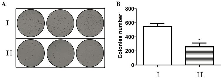
Inhibition Of Mir 155 A Therapeutic Target For Breast Cancer Prevented In Cancer Stem Cell Formation Ios Press

Soft Agar Colony Formation Assay A Representative Pictures Of The Download Scientific Diagram
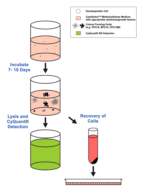
Hematopoietic Cfc Assays
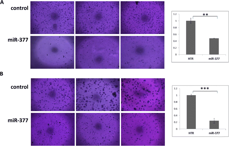
Mir 377 Targets E2f3 And Alters The Nf Kb Signaling Pathway Through Map3k7 In Malignant Melanoma Molecular Cancer Full Text
Onlinelibrary Wiley Com Doi Pdf 10 2164 Jandrol 109

Imagej Macros For The User Friendly Analysis Of Soft Agar And Wound Healing Assays Biotechniques

Colony Formation An Overview Sciencedirect Topics
Http Www Meditecno Pt Upload Product Archive Cba 150 T Tumor Sensitivity Assay Pdf
Plos One Immortalization Of Chicken Preadipocytes By Retroviral Transduction Of Chicken Tert And Tr
Plos One Variable Expression Of Pik3r3 And Pten In Ewing Sarcoma Impacts Oncogenic Phenotypes

Tumor Sensitivity Assay Cell Biolabs Bio Connect

The Soft Agar Colony Formation Assay Protocol
Biomedres Us Pdfs Bjstr Ms Id Pdf
2

Soft Agar Colony Formation Assay As A Hallmark Of

Soft Agar Colony Formation Assay By Creative Bioarray22 Issuu

Cell Based Soft Agar Assays Reaction Biology
Www Mdpi Com 73 4409 8 2 117 Pdf

Suppression Of Mitochondrial Respiration With Local Anesthetic Ropivacaine Targets Breast Cancer Cells Gong Journal Of Thoracic Disease
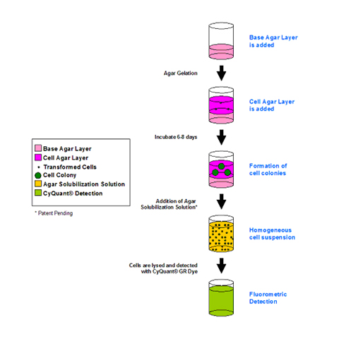
96 Well Cell Transformation Assays Standard Soft Agar

Cytoselect 96 Well In Vitro Tumor Sensitivity Assay Soft Agar Colony Formation Assay Kit
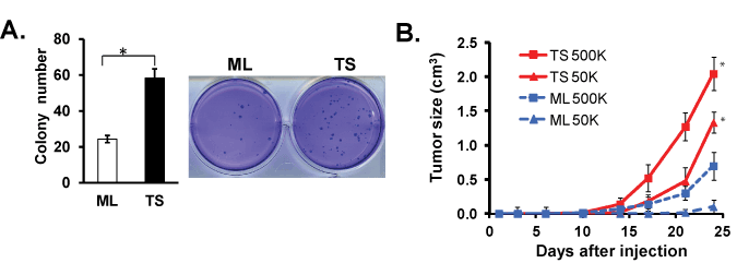
Figure 3
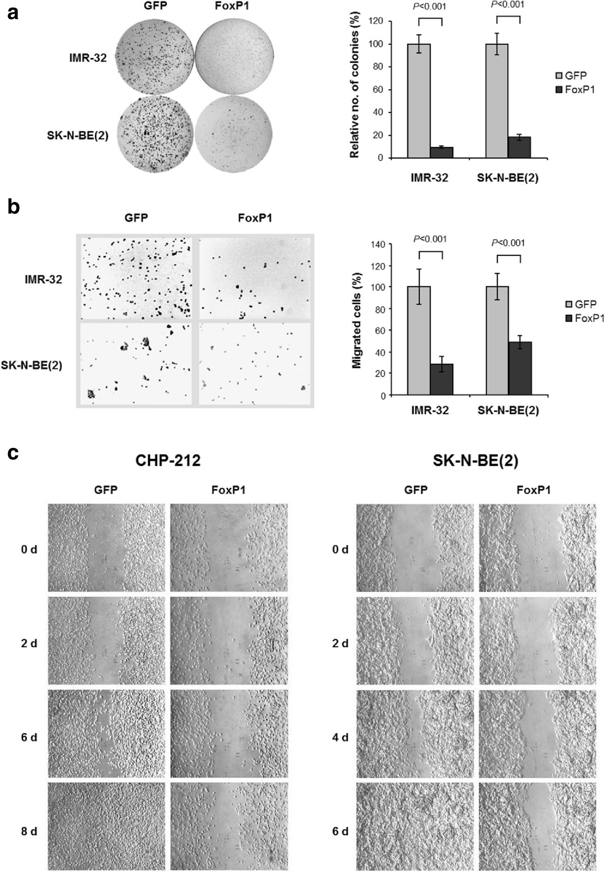
Foxp1 Inhibits Cell Growth And Attenuates Tumorigenicity Of Neuroblastoma Bmc Cancer Full Text
Q Tbn And9gcsqetcsbbvalvrfgtovtcg8ezyad Pvptypsumasbx Zotdygr6 Usqp Cau

Figure 2 Immortalized Cancer Associated Fibroblasts Promote Prostate Cancer Carcinogenesis Proliferation And Invasion Anticancer Research

Representative Soft Agar Colony Formation Assay For The Tvm A12 Download Scientific Diagram

Soft Agar Colony Formation Assay Of Hela Cells Hela Cells Enclosed In Download Scientific Diagram
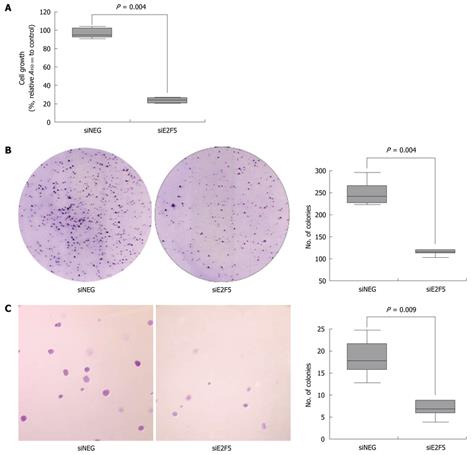
A Potential Oncogenic Role Of The Commonly Observed E2f5 Overexpression In Hepatocellular Carcinoma
Www Bioscience Co Uk Userfiles Pdf Cba 130 Cell Transformation Assay 1 Pdf

Soft Agar Colony Formation Assay Of Hipscs And Teratocarcinoma Pa 1 Download Scientific Diagram

Cytoselect Cell Transformation Assays Cell Biolabs Bio Connect
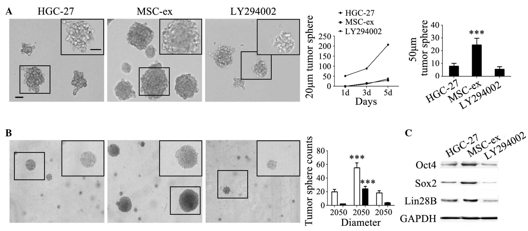
Exosomes Derived From Human Mesenchymal Stem Cells Promote Gastric Cancer Cell Growth And Migration Via The Activation Of The Akt Pathway
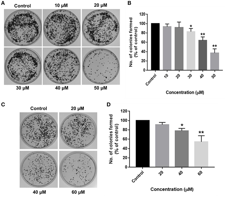
Frontiers Rotundic Acid Induces Dna Damage And Cell Death In Hepatocellular Carcinoma Through Akt Mtor And Mapk Pathways Oncology

Identification Of ox1 As A Therapeutic Target In Triple Negative Breast Cancer Cancer Discovery
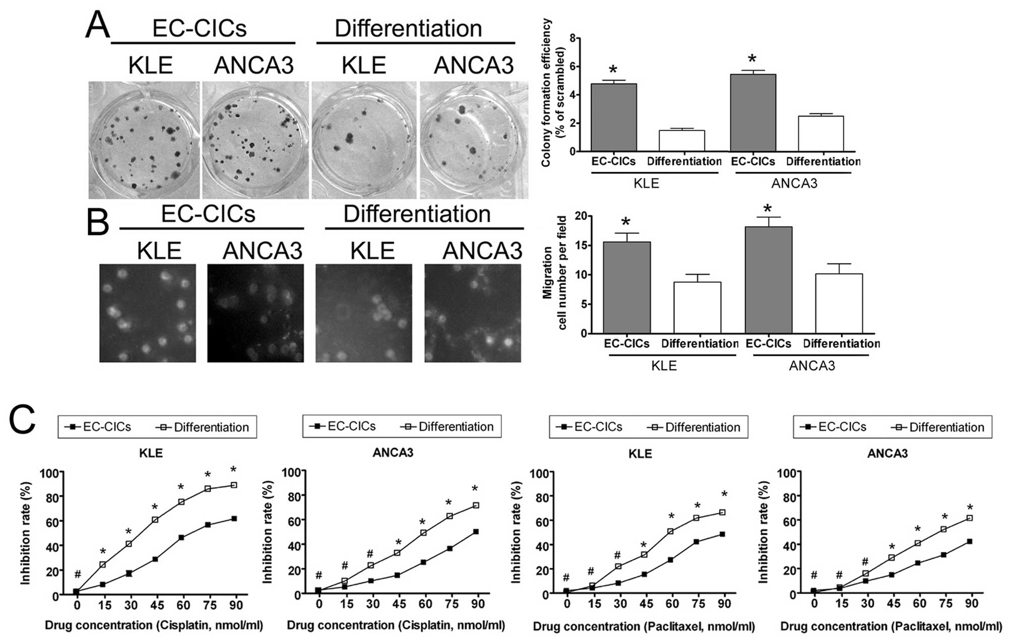
Isolation And Characterization Of Proliferative Migratory And Multidrug Resistant Endometrial Carcinoma Initiating Cells From Human Type Ii Endometrial Carcinoma Cell Lines
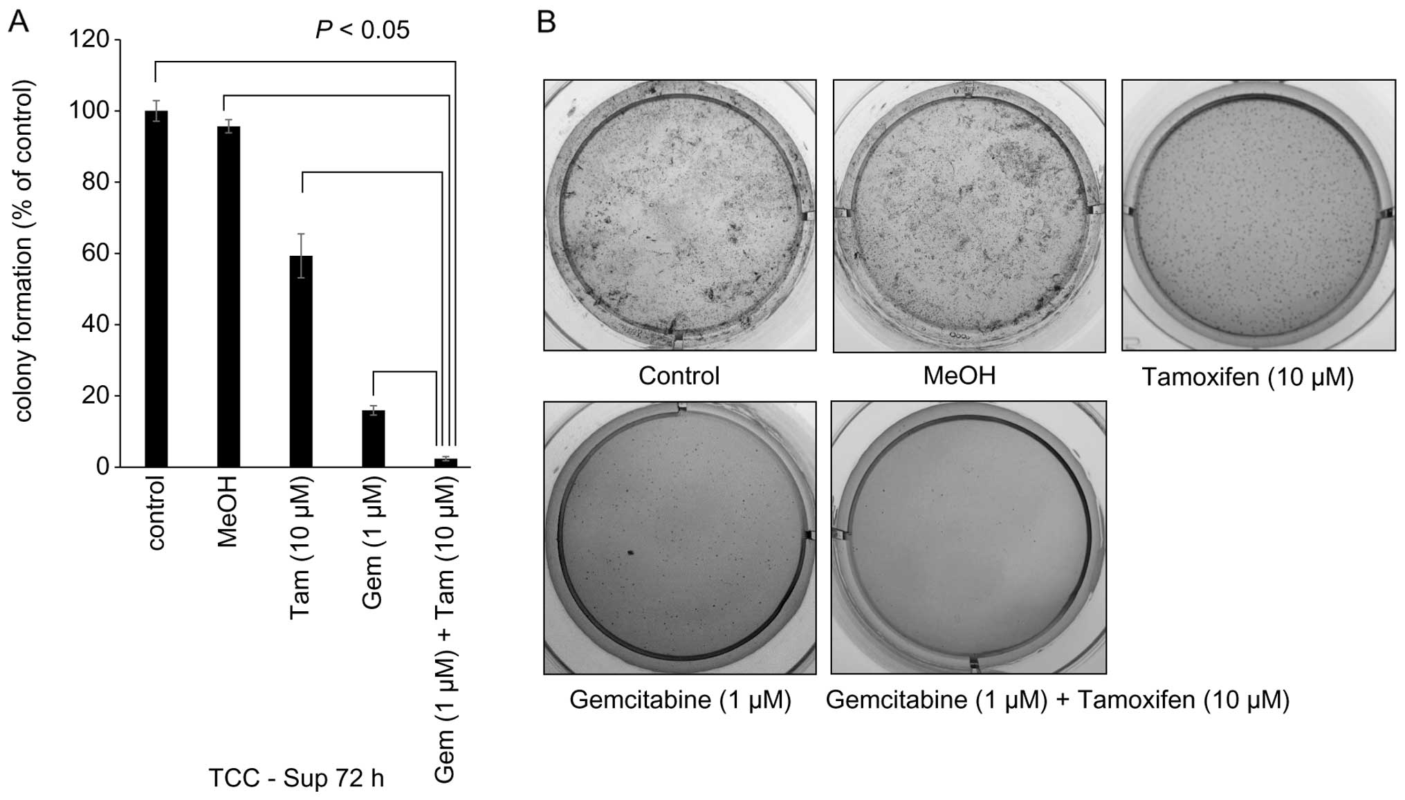
Sequential Gemcitabine And Tamoxifen Treatment Enhances Apoptosis And Blocks Transformation In Bladder Cancer Cells

K921 100 Cell Transformation Assay Kit Colorimetric Clinisciences

Fig 5 Functional Study Of Vim In Siha Cervical Cancer Cells A Soft Agar Colony Forming Assays Siha Cervical Calls Were Transfected With Pcdna3 Or Pcdna3 Vim Vectors And The Transfected Cells Were Plated With 50 Cell Numbers Per 6 Well Plate For 22 Days
Journals Sagepub Com Doi Pdf 10 1177
Http Valterocchiena Com Pdf Listino Cell Biolabs Pdf

Oncogenic Transformation Of Human Mammary Epithelial Cells By Autocrine Human Growth Hormone Cancer Research

Cell Based Soft Agar Assays Reaction Biology

Utilization Of The Soft Agar Colony Formation Assay To Identify Inhibitors Of Tumorigenicity In Breast Cancer Cells Protocol
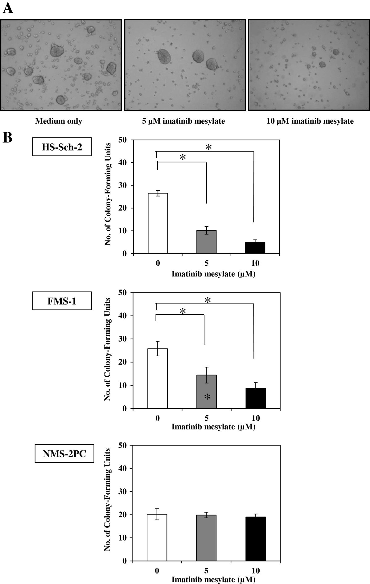
Imatinib Mesylate Inhibits Cell Growth Of Malignant Peripheral Nerve Sheath Tumors In Vitro And In Vivo Through Suppression Of Pdgfr B Bmc Cancer Full Text
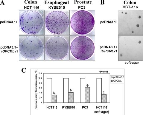
Softagar Soft Agar Www Dingjisc Com
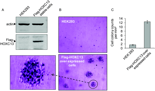
Antisense Oligonucleotide Mediated Knockdown Of Hoxc13 Affects Cell Growth And Induces Apoptosis In Tumor Cells And Over Expression Of Hoxc13 Induces Rsc Advances Rsc Publishing Doi 10 1039 C2ra206g

Full Text Trim31 Promotes Glioma Proliferation And Invasion Through Activating N Ott

Cytoselect 96 Well In Vitro Tumor Sensitivity Assay Soft Agar Colony Formation Assay Kit
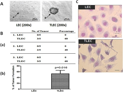
Morphology And Tumorigenic Properties Of Tlecsa Soft A Open I
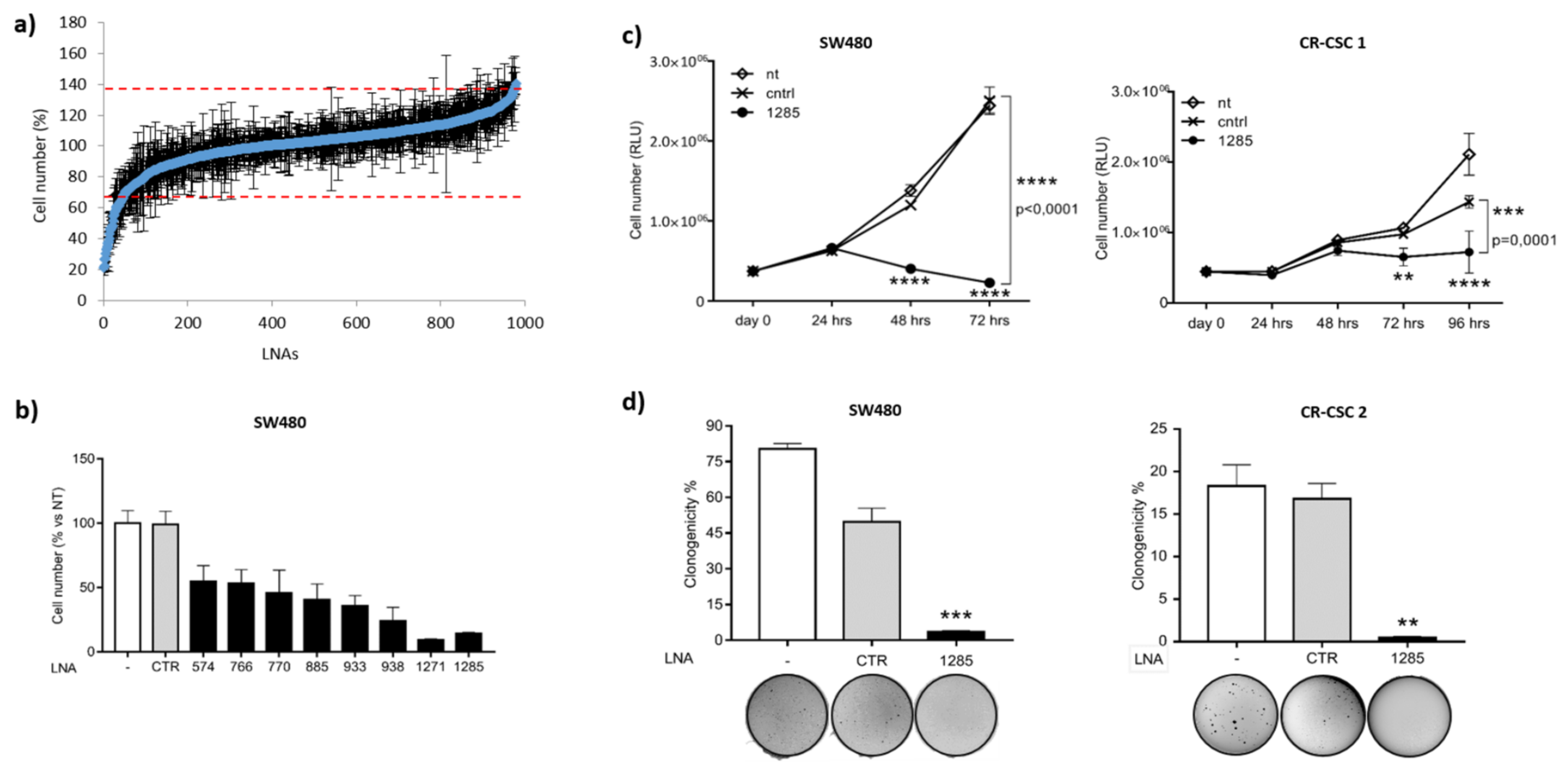
Ijms Free Full Text Mir 1285 3p Controls Colorectal Cancer Proliferation And Escape From Apoptosis Through Dapk2 Html

Oncogenic Transformation Of Human Mammary Epithelial Cells By Autocrine Human Growth Hormone Cancer Research
Www Bionova Es Documents Cellbiolabs Cell Based Assays Pdf

Oxford Optronix Gelcount

The Soft Agar Colony Formation Assay Protocol
Plos One Long Non Coding Rna Hotair Regulates The Proliferation Self Renewal Capacity Tumor Formation And Migration Of The Cancer Stem Like Cell Csc Subpopulation Enriched From Breast Cancer Cells
Artscimedia Case Edu Wp Content Uploads Sites 198 16 10 Soft Agar Assay Protocol Pdf

A Novel Hydrazide Compound Exerts Anti Metastatic Effect Against Breast Cancer
Onlinelibrary Wiley Com Doi Pdf 10 1111 J 1349 7006 12 X
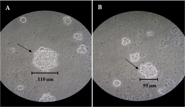
Establishment And Characterization Of Two Human Breast Carcinoma Cell Lines By Spontaneous Immortalization Discordance Between Estrogen Progesterone And Her2 Neu Receptors Of Breast Carcinoma Tissues With Derived Cell Lines Cancer Cell International
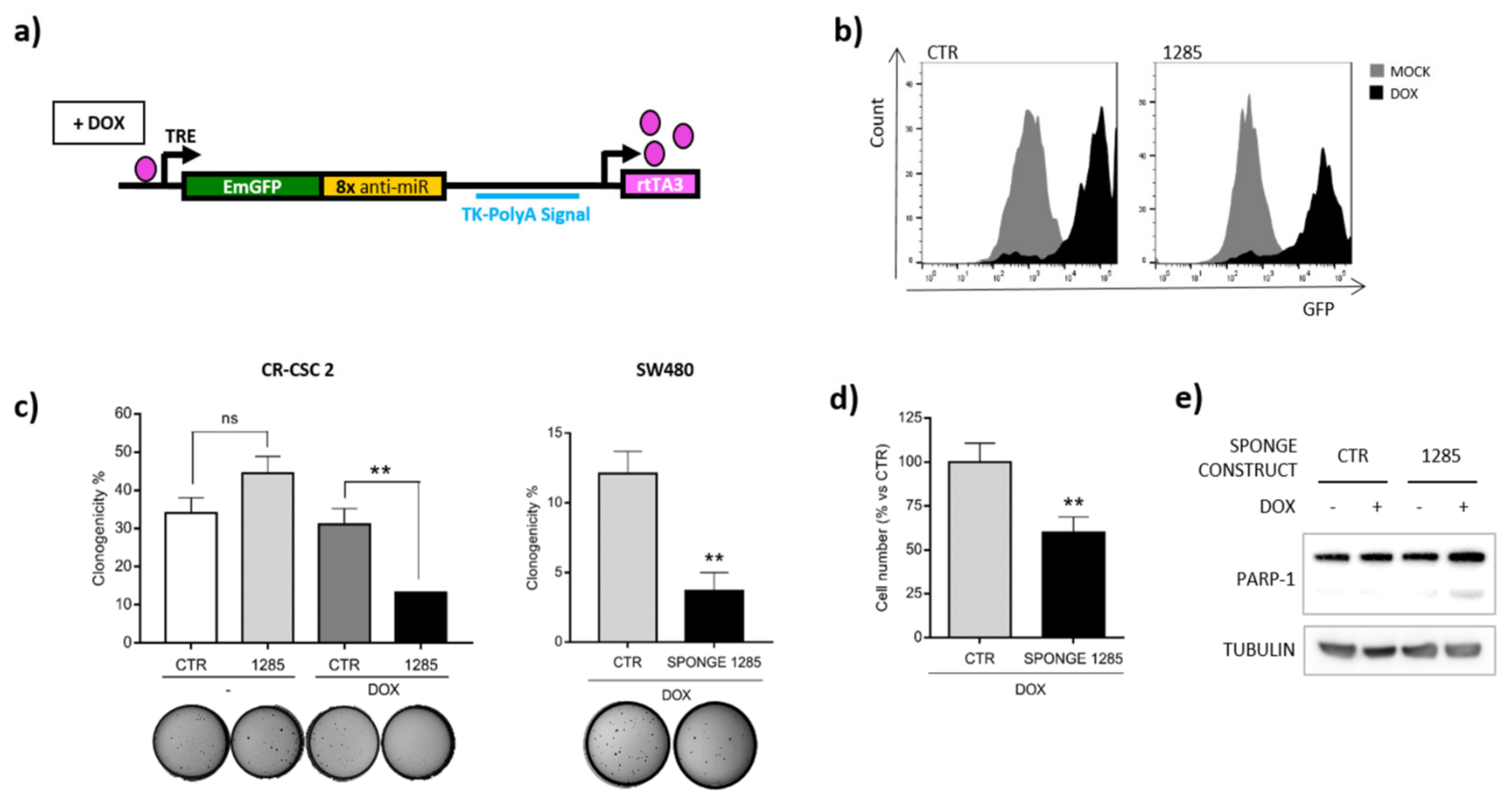
Ijms Free Full Text Mir 1285 3p Controls Colorectal Cancer Proliferation And Escape From Apoptosis Through Dapk2 Html

Imagej Macros For The User Friendly Analysis Of Soft Agar And Wound Healing Assays Biotechniques
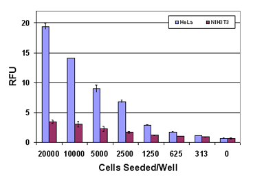
96 Well Cell Transformation Assays Standard Soft Agar

Synergistic Inhibition Of Soft Agar Colony Formation By The Download Scientific Diagram

Jci Afap 110 Is Overexpressed In Prostate Cancer And Contributes To Tumorigenic Growth By Regulating Focal Contacts

Wnt10a Increases Colony Formation Ability
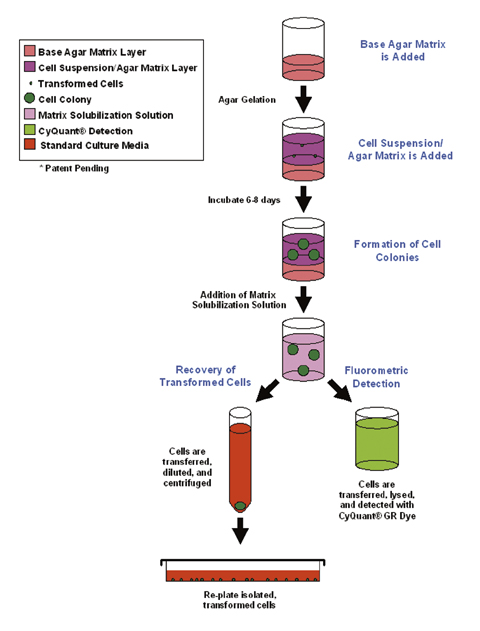
96 Well Cell Transformation Assays Soft Agar With Cell Recovery
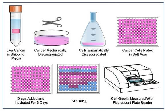
Soft Agar Colony Formation Assay Creative Bioarray Creative Bioarray

Figure 2 From Strebloside Induced Cytotoxicity In Ovarian Cancer Cells Is Mediated Through Cardiac Glycoside Signaling Networks Semantic Scholar

A Rate Of Proliferation Of Pc3 Prostate Cancer Cells Is Greatly Reduced By Treatment With Oligomers Containing Cpg Motifs As Assessed By A Soft Agar Ppt Download
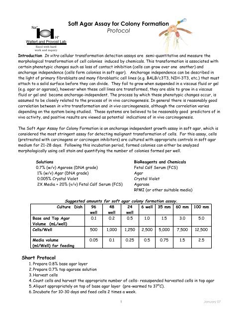
Soft Agar Assay Protocol
Www 2bscientific Com Getmedia 48cf7444 f3 4bbe B1ca Df2df3e965 Colony Formation Brochure 14 Pdf

Utilization Of The Soft Agar Colony Formation Assay To Identify Inhibitors Of Tumorigenicity In Breast Cancer Cells Abstract Europe Pmc

A Colony Formation Assay Shows A Concentration Dependent Decrease In Download Scientific Diagram
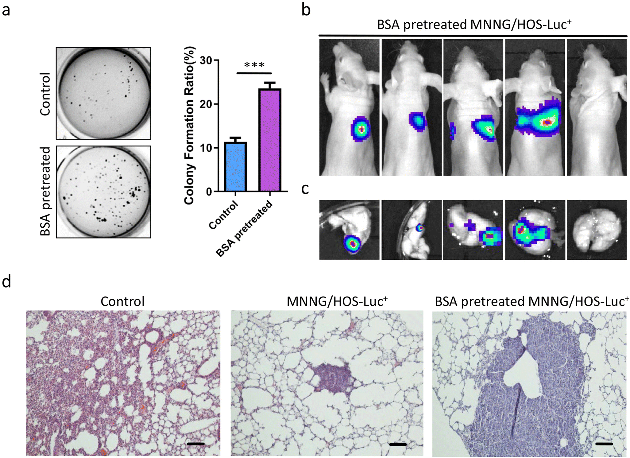
Figure 3 Interstitial Serum Albumin Empowers Osteosarcoma Cells With Faim2 Transcription To Obtain Viability Via Dedifferentiation Springerlink

Pdf The Soft Agar Colony Formation Assay Semantic Scholar

Refractory Nature Of Normal Human Diploid Fibroblasts With Respect To Oncogene Mediated Transformation Pnas

Utilization Of The Soft Agar Colony Formation Assay To Identify Inhibitors Of Tumorigenicity In Breast Cancer Cells Protocol

Expression Of Q227l Gas In Mcf 7 Human Breast Cancer Cells Inhibits Tumorigenesis Pnas
Indirect Soft Agar Colony Formation Assay With Hela Cells And Download Scientific Diagram
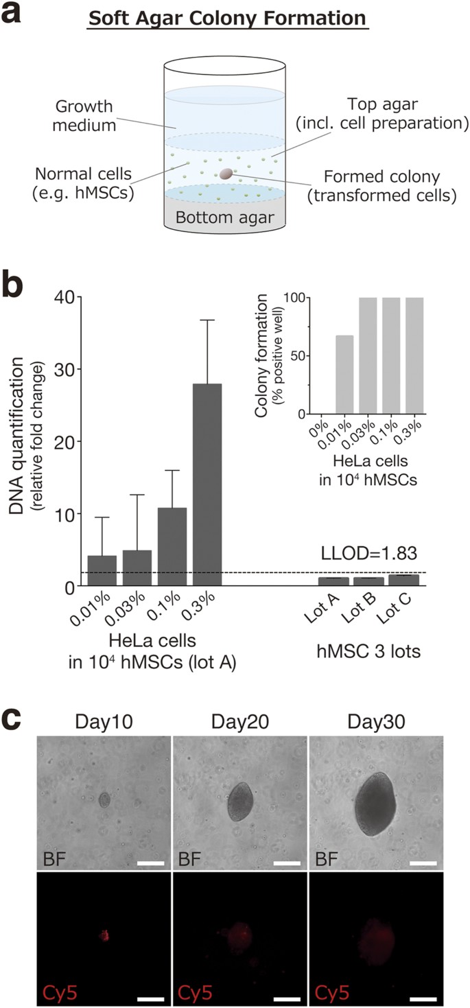
Ultra Sensitive Detection Of Tumorigenic Cellular Impurities In Human Cell Processed Therapeutic Products By Digital Analysis Of Soft Agar Colony Formation Scientific Reports

A Colony Formation Assay In Ags Cells B Soft Agar Colony Formation Download Scientific Diagram

Soft Agar Assay With Temperature Responsive Poly 2 3 Glucose Gel

Leonid Schneider Visit My Site For Covid19 Cures A Twitter Smutclyde A Reviewer Asked For Representative Pictures For Transwell Migration And Soft Agar Colony Formation Assays And These Were Duly Added In The
Http Ar Iiarjournals Org Content 24 6 39 Full Pdf

Full Text Shp2 Overexpression Enhances The Invasion And Metastasis Of Ovarian Ca Ott
Biopply Com Assets References Cfa Abc Biopply Pdf

Figure 1
Www Bionova Es Documents Cellbiolabs Cell Based Assays Pdf

Catalogo Cell Biolabs By Fermelo Biotec Issuu
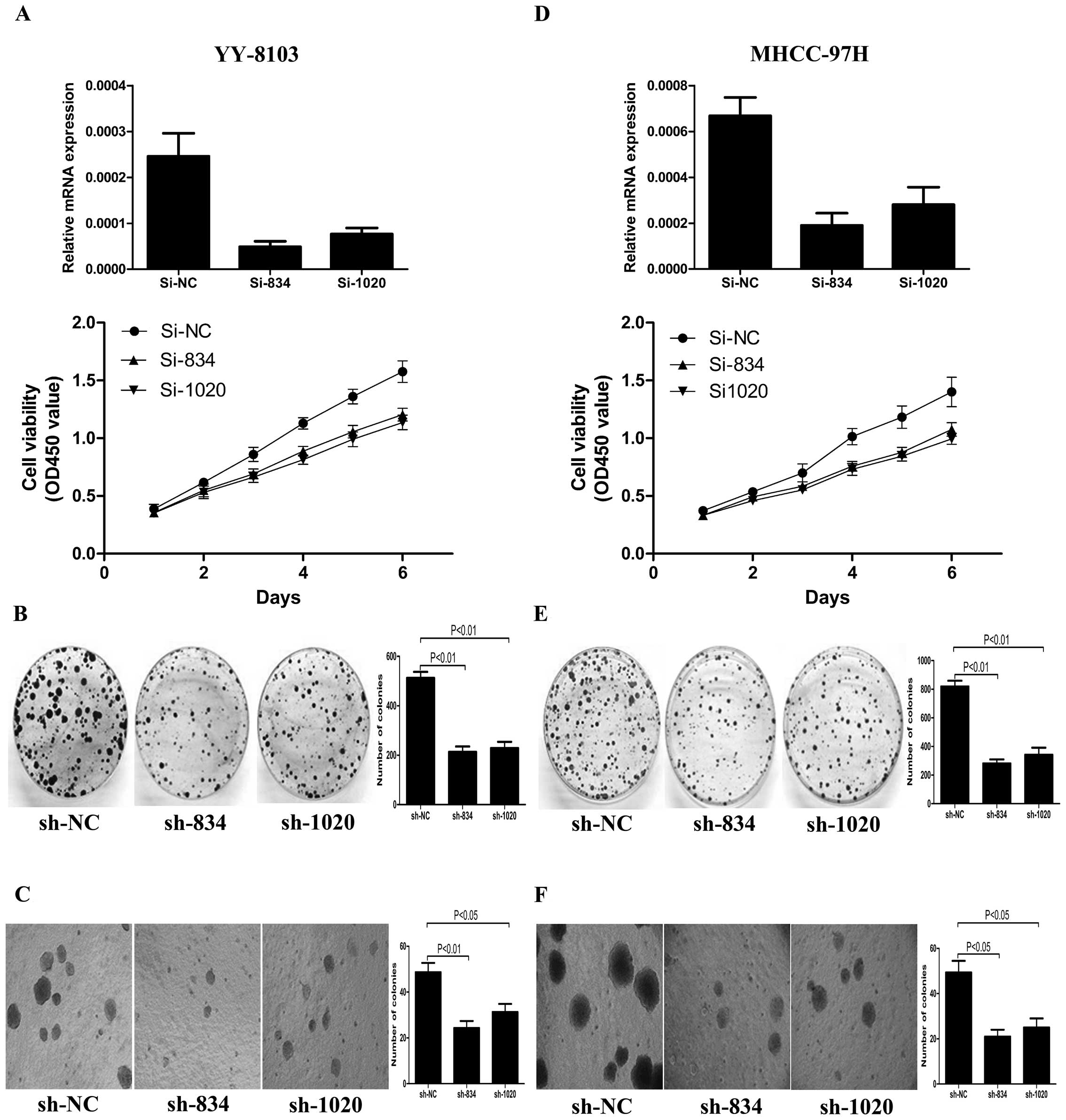
Cdca7l Promotes Hepatocellular Carcinoma Progression By Regulating The Cell Cycle

The Effects Of Me2 On Soft Agar Colony Formation In Human Glioma Cell Download Scientific Diagram
Plos One Targeting Insulin Like Growth Factor 1 Receptor Inhibits Pancreatic Cancer Growth And Metastasis

Figure 1 From Automated Soft Agar Assay For The High Throughput Screening Of Anticancer Compounds Semantic Scholar
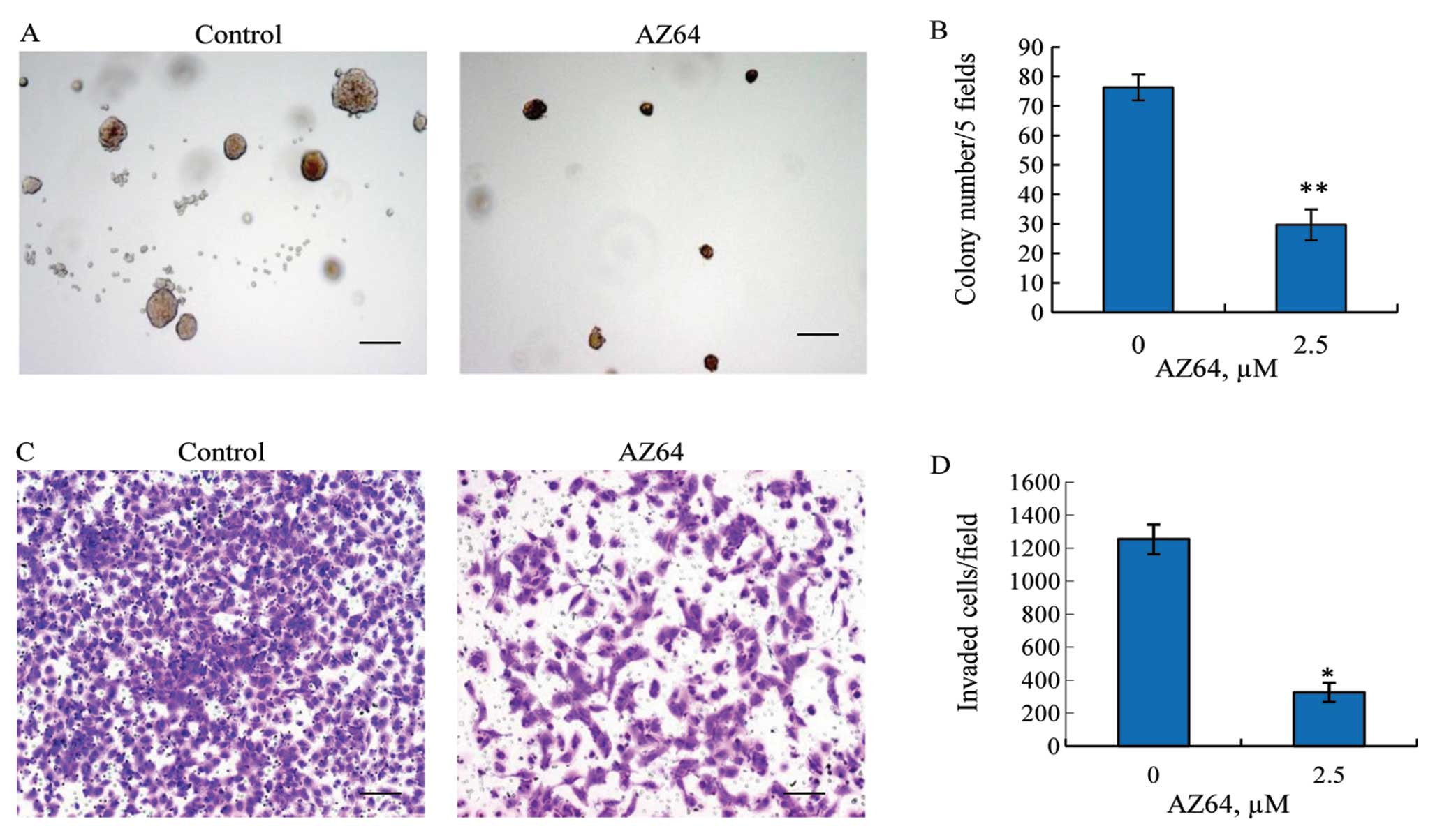
Antitumor Activity Of Az64 Via G2 M Arrest In Non Small Cell Lung Cancer

Proliferation And Invasion Plasticity In Tumor Cells Pnas


