Mrt Cranial
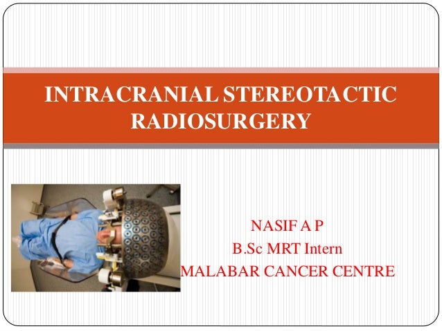
Intracranial Stereotactic Radiosurgery
T2 Weighted Mrt In Coronar Projection Of A Woman With Status After Download Scientific Diagram
Tumor Simulierende Lasionen In Der Kranialen Mrt
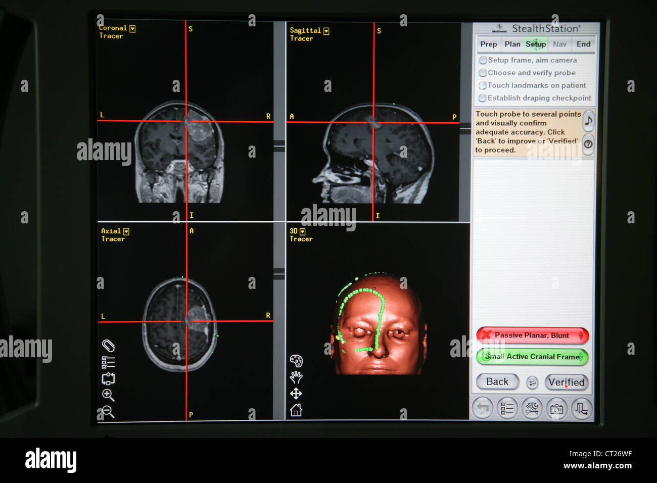
Meningeom Mrt Stockfotografie Alamy

Mri Images Of Brain With Stroke Mri Scan Images Kopfschmerzen Gehirn Overnight Oats Gesund

Animal Mrt Cruciate Ligament Tear In Dogs Cruciate Ligament In Dogs Cruciate Ligament Cruciate Ligament Tear
This cover page design template is complete compatible with Google Docs Just download DOCX format and open the theme in Google Docs Unfortunately, the item MRT Of Cranial Cavity Word Template id which price is Free has no available description, yet The item rating has 46 star(s) with 23 votes.
Mrt cranial. Describe one patient with Miller Fisher syndrome (MFS) and another with cranial neuropathies Both developed ocular motility deficits after a viral prodrome of fever, anosmia, and ageusia The patient with MFS had elevated CSF protein, positive serum antiGD1b ganglioside antibodies, and a swift therapeutic response to IV immunoglobulin (IVIg). Background Cranial ultrasound becomes an important diagnostic tool to evaluate brain injury in infants Brain injury is a major complication for preterm birth The brain injury of preterm infants. An atypical teratoid rhabdoid tumor (AT/RT) is a rare tumor usually diagnosed in childhood Although usually a brain tumor, AT/RT can occur anywhere in the central nervous system (CNS), including the spinal cord About 60% will be in the posterior cranial fossa (particularly the cerebellum).
MRI Atlas of the Brain This page presents a comprehensive series of labeled axial, sagittal and coronal images from a normal human brain magnetic resonance imaging exam This MRI brain crosssectional anatomy tool serves as a reference atlas to guide radiologists and researchers in the accurate identification of the brain structures. MRI of the head;. Cranial MRI Definition An MRI of the head is a noninvasive procedure that uses powerful magnets and radio waves to construct pictures of the clear, detailed pictures of brain tissues.
Five DNA Methylation Subgroups of RTs from Cranial and ExtraCranial Sites Correlate with Previously Known ATRT and MRT Subgroups, Anatomical Sites, and SMARCB1 Deletion Patterns (A)UnsupervisedNMFanalysiswasperformedusingthetop10,000mostvariablymethylatedCpGsitesandrevealedfivesubgroups(top)Clinicalfeatures,gene expression subgroups of MRTs, and previously characterized ATRT subgroups are shown in colored tracks (middle). New MRT Imaging Biomarkers and Treatment With Kinetic Oscillatory Stimulation (KOS) in Nasal Cavity for Myalgic Encephalomyelitis/Chronic Fatigue Syndrome (ME/CFS) The safety and scientific validity of this study is the responsibility of the study sponsor and investigators. Cranial MR imaging performed at the time of admission with a 15T superconducting magnet showed small cortical veins and superior sagittal, straight, transverse, and sigmoid sinuses but no intracranial mass lesion, ventricular dilation, or sinus thrombosis (Fig 1A).
The brainstem is the distal part of the brain that extends from the base of the brain to the spinal cord From superior to inferior, the brainstem consists of the midbrain, pons and medulla oblongata Each of them features many important structures such as the cranial nerve nuclei. Brain imaging, magnetic resonance imaging of the head or skull, cranial magnetic resonance tomography (MRT), neurological MRI they describe all the same radiological imaging technique for medical. Hardbound MRI Textbook MRI BIOEFFECTS, SAFETY, AND PATIENT MANAGEMENT is a comprehensive, authoritative textbook on the health and safety concerns of MRI technology that contains contributions from more than forty internationally respected experts in the field It serves as the definitive resource for radiologists and other physicians, MRI technologists, physicists, scientists, MRI facility.
A PET scan is an imaging exam that’s used to diagnose diseases or issues by looking at how the body is functioning It uses a special dye with radioactive tracers to help the machine capture. This section of the website will explain large and minute details of arterial anatomy of brain. An MRI (magnetic resonance imaging) lets your doctor see the organs, bones, and tissues inside your body without having to do surgery This test can help diagnose a disease or injury You might.
Magnetic Resonance Imaging (MRI) is a commonly accepted and widely used diagnostic medical procedure It is often safe to perform MRI on an individual that has an orthopaedic implant device. Overview Whiplash is a neck injury due to forceful, rapid backandforth movement of the neck, like the cracking of a whip Whiplash is commonly caused by rearend car accidents. The UPRIGHT MRI has demonstrated pronounced anatomic pathology of the cervical spine in five of the MS patients studied and definitive cervical pathology in the other three The pathology was the result of prior head and neck trauma.
Cavernous malformations (CMs) of the brain are vascular malformations with an estimated prevalence between 04 and 09% 1 , appearing mainly as singular supratentorial lesions 2 These lesions are made up of clusters of deformed vessels, lined by endothelium, and filled with blood at various stages of thrombosis. CranioCurve® Preformed Mesh CranioCurve ® Preformed Mesh CranioCurve Preformed Mesh is a titanium cranioplasty solution and is part of our comprehensive cranioplasty portfolio, for efficient coverage of cranial defects in multiple anatomic regions. Malignant rhabdoid tumors (MRTs) are rare lethal tumors of childhood that most commonly occur in the kidney and brain MRTs are driven by SMARCB1 loss, but the molecular consequences of SMARCB1.
Malignant rhabdoid tumors (MRTs) are rare lethal tumors of childhood that most commonly occur in the kid ney and brain MRTs are driven by SMARCB1 loss, but the molecular consequences of SMARCB1 loss in extracranial tumors have not been comprehensively described and genomic resources for analyses of extracranial MRT are limited. Rhaboid tumors that grow outside of the brain are called extracranial malignant rhabdoid tumor, malignant rhabdoid tumor, or MRT MRTs grow and spread to other parts of the body quickly How common are extracranial malignant rhabdoid tumors?. Was bedeutet cranial in einem MRTBefund vom Kopf?.
PURPOSE Malignant rhabdoid tumor (MRT) is a rare and highly aggressive tumor that affects young children Due to its extreme rarity, most of the available data are based on retrospective case series To add to the current knowledge of this disease, we reviewed the patients treated for extracranial MRT in our institute. Background Cranial ultrasound becomes an important diagnostic tool to evaluate brain injury in infants Brain injury is a major complication for preterm birth The brain injury of preterm infants. Magnetic resonance imaging (MRI) is a safe, painless test that uses radio waves and energy from strong magnets to create detailed images of your body A cervical MRI scans the soft tissues of your.
The ninth cranial nerve, which exits the skull through the jugular foramen, has both motor and sensory components Cell bodies of motor neurons, located in the nucleus ambiguus in the medulla oblongata, project as special visceral efferent fibres to the stylopharyngeal muscle. Hier erklären Ärzte leicht verständlich Begriffe aus medizinischen Befunden. Magnetic resonance imaging cranial;.
MRI is the investigative tool of choice for neurological cancers over CT, as it offers better visualization of the posterior cranial fossa, containing the brainstem and the cerebellum. Magnetic resonance angiography – also called a magnetic resonance angiogram or MRA – is a type of MRI that looks specifically at the body’s blood vessels. MRI with contrast enhancement is a valuable tool for detecting and characterizing disease of the cranial nerves Abnormal cranial nerve enhancement on MRI may sometimes be the first or only indication of an underlying disease process.
The complex anatomy of the head and neck involves many vital structures, such as cranial nerves and important blood vessels The MRT enables doctors to plan a safe approach to a lesion in this area without endangering critical nerves and arteries. CNS = brain, brain meninges, cranial nerves & spinal cord PNS = nerves and ganglia Has both sensory and motor function Sensory send impulses to brain and spinal cord to skeletal muscle Autonomic conducts impulses from brain and spinal cord to smooth muscle tissue Ex Digestive and cardiopulmonary systems. Which settle in the mesenteric veins (5) The adultwormlayeggsthatareexcretedwithstool or urine Different mechanisms of invasion of thebrainhavebeendiscussedtheeggsmay.
The twelve cranial nerves consist of the olfactory (I), optic (II), oculomotor (III), trochlear (IV), trigeminal (V), abducens (VI), facial (VII), vestibulocochlear (VIII), glossopharyngeal (IX), vagus (X), accessory (XI), and hypoglossal (XII) nerves Each nerve has an intraaxial, cisternal, dural, osseous, and extracranial segment. The use of MRT and MT appeared to be effective in individuals with working mechanical NP, although MRT seemed to be a more beneficial intervention therapy for the craniovertebral angle, active cervical ranges of motion (side bending and rotation), and QoL (Mental Component Summary and Social Function dimension) 17 In the original study, it would be expected that the favorable results regarding craniovertebral angle and active cervical ranges of motion were in line with the results regarding. This module of human anatomy is useful for residents and students who wish to learn the basics of the anatomy of the cervical spine in MRI on a 15 Tesla device It was created from sagittal T1weighted sequences and T2 reconstructions on the three reference planes (sagittal, coronal and axial) with 1mm slices.
The conclusion of a recent large cohort study from Ontario, Canada (Ray JG et al JAMA 16;316(9)) states, "Exposure to MRI during the first trimester of pregnancy compared with nonexposure was not associated with increased risk of harm to the fetus or in early childhood Gadolinium MRI at any time during pregnancy was associated with an increased risk of a broad set of. Hardbound MRI Textbook MRI BIOEFFECTS, SAFETY, AND PATIENT MANAGEMENT is a comprehensive, authoritative textbook on the health and safety concerns of MRI technology that contains contributions from more than forty internationally respected experts in the field It serves as the definitive resource for radiologists and other physicians, MRI technologists, physicists, scientists, MRI facility. MRI with contrast enhancement is a valuable tool for detecting and characterizing disease of the cranial nerves Abnormal cranial nerve enhancement on MRI may sometimes be the first or only indication of an underlying disease process Address correspondence to F Saremi (email protected).
Magnetic resonance imaging (MRI) is a medical imaging technique used in radiology to form pictures of the anatomy and the physiological processes of the body MRI scanners use strong magnetic fields, magnetic field gradients, and radio waves to generate images of the organs in the body MRI does not involve Xrays or the use of ionizing radiation, which distinguishes it from CT and PET scans. Cranial CT Scan Medically reviewed by Deborah Weatherspoon, PhD, RN, CRNA A cranial CT scan of the head is a diagnostic tool used to create detailed pictures of the skull, brain, paranasal. The use of MRT and MT appeared to be effective in individuals with working mechanical NP, although MRT seemed to be a more beneficial intervention therapy for the craniovertebral angle, active cervical ranges of motion (side bending and rotation), and QoL (Mental Component Summary and Social Function dimension) 17 In the original study, it would be expected that the favorable results regarding craniovertebral angle and active cervical ranges of motion were in line with the results regarding.
Molecular radiotherapy (MRT) with Iodine131 metaiodobenzylguanidine (131ImIBG), a noradrenaline analogue taken up by neuroblastomas, phaeochromocytoma and paragangliomas that overexpress the noradrenaline transporter, has long been used for the treatment of refractory or relapsed neuroblastoma, and has also been incorporated into induction and consolidation regimens. An MRI is a test your doctor can use to diagnose and monitor different conditions Find out why you might need this test and how it works. The UPRIGHT MRI has demonstrated pronounced anatomic pathology of the cervical spine in five of the MS patients studied and definitive cervical pathology in the other three The pathology was the result of prior head and neck trauma.
The complex anatomy of the head and neck involves many vital structures, such as cranial nerves and important blood vessels The MRT enables doctors to plan a safe approach to a lesion in this area without endangering critical nerves and arteries. This cover page design template is complete compatible with Google Docs Just download DOCX format and open the theme in Google Docs Unfortunately, the item MRT Of Cranial Cavity Word Template id which price is Free has no available description, yet The item rating has 46 star(s) with 23 votes. ADVERTISEMENT Radiopaedia is free thanks to our supporters and advertisers Become a Gold Supporter and see no ads.
This site provides clear and easily accessible guide to many of the practical aspects of MRI including MRI protocols, MRI planning, MRI anatomy, MRI techniques, MRI safety and much more. A new high‐resolution MR protocol for the combined assessment of neurovascular arterial anatomy and subsequent evaluation of inflammatory disease in cranial vessels walls has been investigated. MR Safe – MR Conditional Trauma DePuy Synthes 3 MR TERMS The new MR safe will be a much higher standard than currently and will be absolute To obtain the new MR safe designation, objects must be completely free of all metallic.
Five DNA Methylation Subgroups of RTs from Cranial and ExtraCranial Sites Correlate with Previously Known ATRT and MRT Subgroups, Anatomical Sites, and SMARCB1 Deletion Patterns (A)UnsupervisedNMFanalysiswasperformedusingthetop10,000mostvariablymethylatedCpGsitesandrevealedfivesubgroups(top)Clinicalfeatures,gene expression subgroups of MRTs, and previously characterized ATRT subgroups are shown in colored tracks (middle). Malignant Rhabdoid Tumour (MRT) is an aggressive sarcomatous neoplasm usually arising in the kidney, with a histology distinct from Wilms' tumour Occasionally MRT may arise in other parts of the body, including the brain. In first year MBBS, we all have read cranial nerves anatomy How to apply that knowledge to identify cranial nerves on MRI, is what this video explains in a.
Malignant rhabdoid tumors (MRTs) are rare lethal tumors of childhood that most commonly occur in the kidney and brain MRTs are driven by SMARCB1 loss, but the molecular consequences of SMARCB1 loss in extracranial tumors have not been comprehensively described and genomic resources for analyses of extracranial MRT are limited. MRT is very rare. This MRI cranial nerves axial cross sectional anatomy tool is absolutely free to use Use the mouse scroll wheel to move the images up and down alternatively use the tiny arrows (>>) on both side of the image to move the images>>) on both side of the image to move the images.
Twentytwo magnetic resonance imaging (MRI) brain studies of different breeds of dogs were reviewed to assess the anatomy of cranial nerve (CN) origins and associated skull foramina These included five anatomic studies of normal brains using 2mmthick slices and 17 studies using conventional clini. Brain imaging, magnetic resonance imaging of the head or skull, cranial magnetic resonance tomography (MRT), neurological MRI they describe all the same radiological imaging technique for medical. Cranial MR imaging performed at the time of admission with a 15T superconducting magnet showed small cortical veins and superior sagittal, straight, transverse, and sigmoid sinuses but no intracranial mass lesion, ventricular dilation, or sinus thrombosis (Fig 1A).
Brain imaging, magnetic resonance imaging of the head or skull, cranial magnetic resonance tomography (MRT), neurological MRI they describe all the same radiological imaging technique for medical. The use of MRT and MT appeared to be effective in individuals with working mechanical NP, although MRT seemed to be a more beneficial intervention therapy for the craniovertebral angle, active cervical ranges of motion (side bending and rotation), and QoL (Mental Component Summary and Social Function dimension) 17 In the original study, it would be expected that the favorable results regarding craniovertebral angle and active cervical ranges of motion were in line with the results regarding. Nuclear magnetic resonance cranial;.
The craniocervical junction (CCJ) is comprised of the inferior surface of the skull, the atlas and axis, as well as muscles and connective tissues that attach the skull to the cervical spine The CCJ encloses the central nervous system (CNS), encephalic vasculature and the cerebrospinal fluid (CSF) system The CCJ spans the brainstem to the spinal cord, including the vascular system as well as.

Cranial Base Surgery Neurochirurgie Luzern Prof Dr Med A Sepehrnia
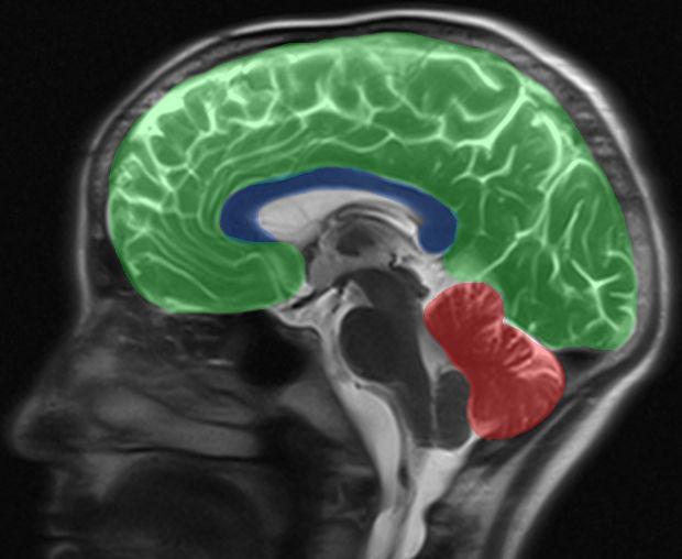
Mrt Des Kopfes

Mrt System Magnetom Prisma Forschungsinfrastruktur

Virtual Screening Based Discovery And Mechanistic Characterization Of The Acylthiourea Mrt 10 Family As Smoothened Antagonists Molecular Pharmacology

The Mrt Amp Leap Protocol For Migraine Special Migraine Special Leaping
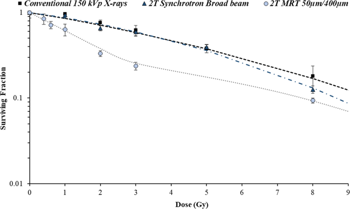
Toward Personalized Synchrotron Microbeam Radiation Therapy Scientific Reports

Identification And Analyses Of Extra Cranial And Cranial Rhabdoid Tumor Molecular Subgroups Reveal Tumors With Cytotoxic T Cell Infiltration Sciencedirect
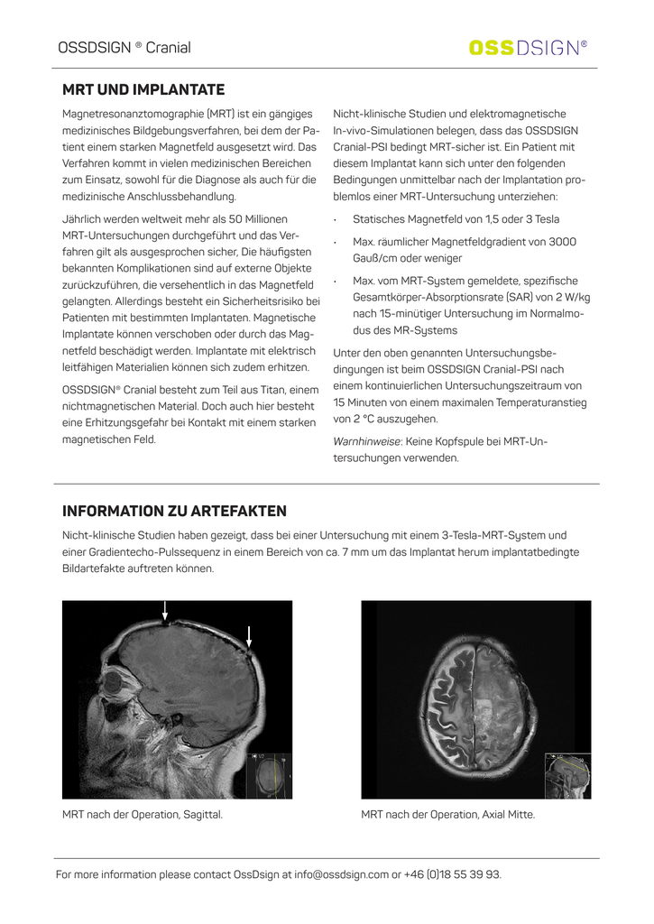
Mrt Und Implantate Information Zu
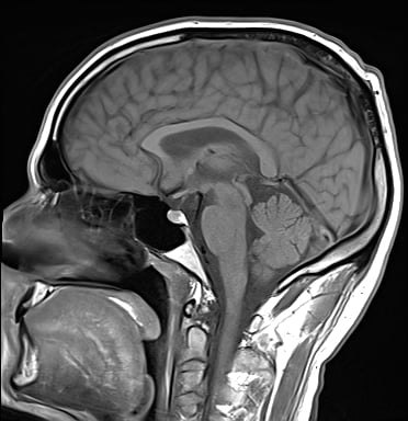
Kopf Mrt Ursachen Fur Kopfschmerzen Schwindel Und Druckgefuhl Finden
Mrt Scan Of The Cranium Arrow Tumorous Mass In The Left Cellulae Download Scientific Diagram

Pdf Mri Based Reevaluation Of Patients With Disc Displacement Without Reduction Mrt Gestutzte Nachuntersuchung Bei Diskusverlagerung Ohne Reposition Semantic Scholar
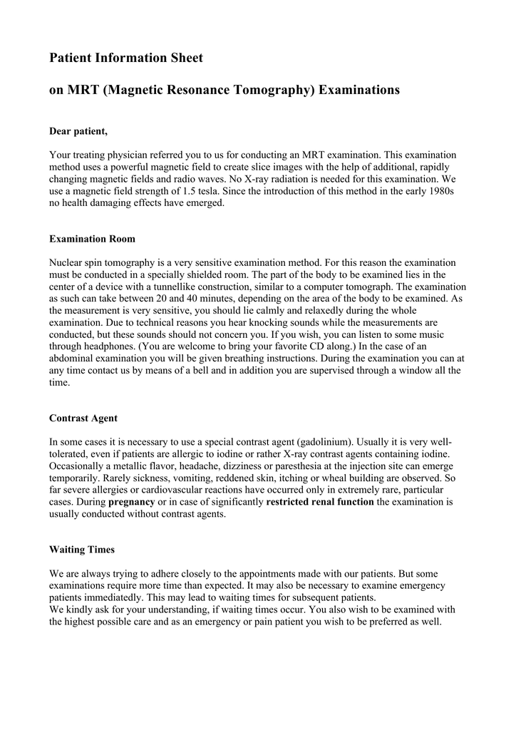
Patient Information Sheet On Mrt Magnetic Resonance

Identification And Analyses Of Extra Cranial And Cranial Rhabdoid Tumor Molecular Subgroups Reveal Tumors With Cytotoxic T Cell Infiltration Sciencedirect

Gadolinium Kontrastmittel Fur Mrt Aufnahme Kann Giftig Sein Welt
Mrt Of Cranial Cavity Word Template 092 Poweredtemplate Com

Mrt System Magnetom Prisma Forschungsinfrastruktur

Identification And Analyses Of Extra Cranial And Cranial Rhabdoid Tumor Molecular Subgroups Reveal Tumors With Cytotoxic T Cell Infiltration Sciencedirect

Funktionelle Mrt Des

Dz10 Mrt Magnetic Resonance Tomgraphy Diagnosezentrum Wien Favoriten

Mrt Kopf Grunde Ablauf Dauer Kosten Praktischarzt

Flair Doccheck Flexikon
Http Ar Iiarjournals Org Content 40 11 6159 Full Pdf

Ms Diagnose Was Zeigt Eine Mrt Untersuchung An

Nuklearmedizin Braunschweig Celler Strasse 30 Braunschweig Mrt Des Kopfes

Her 2 Expression In Four Mrtcell Lines And Two Mrtclinical Tissues Download Scientific Diagram

Mrt Des Kopfes
Www Cell Com Cancer Cell Pdfextended S1535 6108 16 5

Cranial Magnetic Resonance Tomography Mrt From Present Patient A T2 Download Scientific Diagram

Kopf Mrt Ursachen Fur Kopfschmerzen Schwindel Und Druckgefuhl Finden

Imagerie Par Resonance Magnetique Irm Radiologie Baden Baden

Mrt Kopf Grunde Ablauf Dauer Kosten Praktischarzt
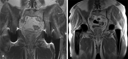
Mrt Diagnostik Von Meniskuslasionen Springerlink
Q Tbn And9gcsbqfnblmoqkflobgztej3cq0tokmgaufnwyhbmjxycvjdd09sb Usqp Cau

Erfahrungsbericht Mrt Mit Platzangst Eine Reportage Vitanet De

Was Passiert Bei Einer Mrt Untersuchung Kinder Erklaren Fur Schulkinder Youtube

Neuroradiologie Wie Behandelbare Formen Der Demenz Erkannt Werden Konnen

Identification And Analyses Of Extra Cranial And Cranial Rhabdoid Tumor Molecular Subgroups Reveal Tumors With Cytotoxic T Cell Infiltration Sciencedirect
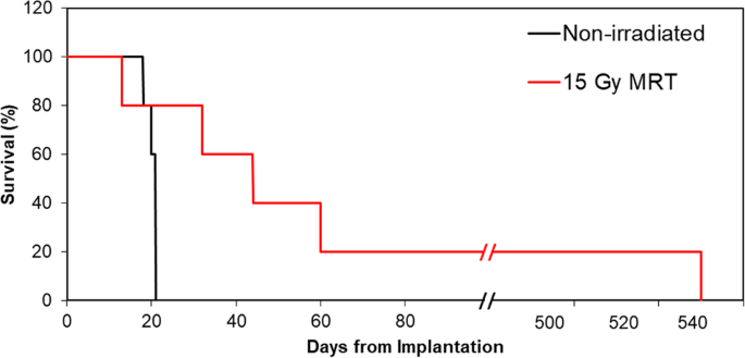
Toward Personalized Synchrotron Microbeam Radiation Therapy Scientific Reports
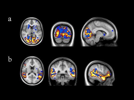
Magnetresonanztomographie Mrt In Der Depressionsforschung 12 Wiley Analytical Science

Clinical Cases Fonar Upright Mri

Gadoliniumhaltige Kontrastmittel Schadlich Fur Das Gehirn

Echtzeit Mrt Film Sprechen Youtube
Mrt Of Cranial Cavity Presentation Template For Powerpoint And Keynote Ppt Star

Novel Two Mrt Cell Lines Established From Multiple Sites Of A Synchronous Mrt Patient Anticancer Research

Angiografie Wikipedia

Craniale Computertomographie Wikipedia
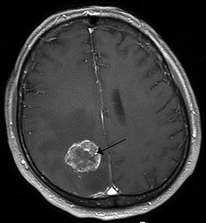
Brain Tumor Wikipedia

Mrt Cell Lines Are Sensitive To Pharmacological Fgfr Inhibition A Download Scientific Diagram
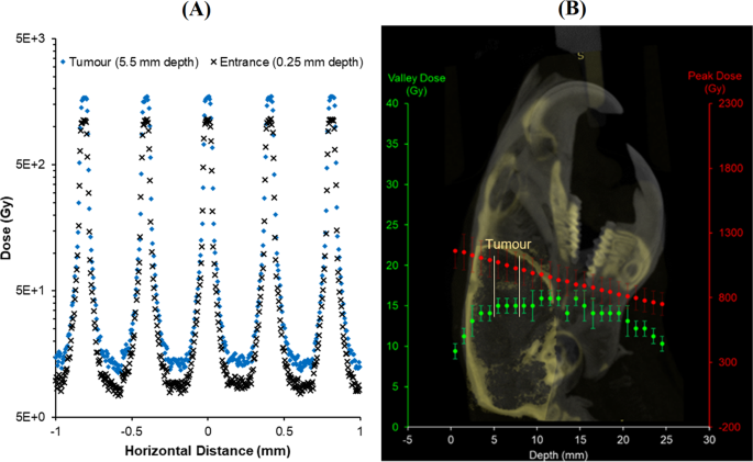
Toward Personalized Synchrotron Microbeam Radiation Therapy Scientific Reports

Clinical Cases Fonar Upright Mri
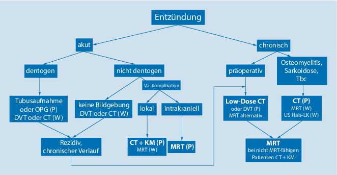
Bildgebung Der Nasennebenhohlen Und Der Frontobasis Springerlink

Nuklearmedizin Braunschweig Celler Strasse 30 Braunschweig Mrt Des Kopfes
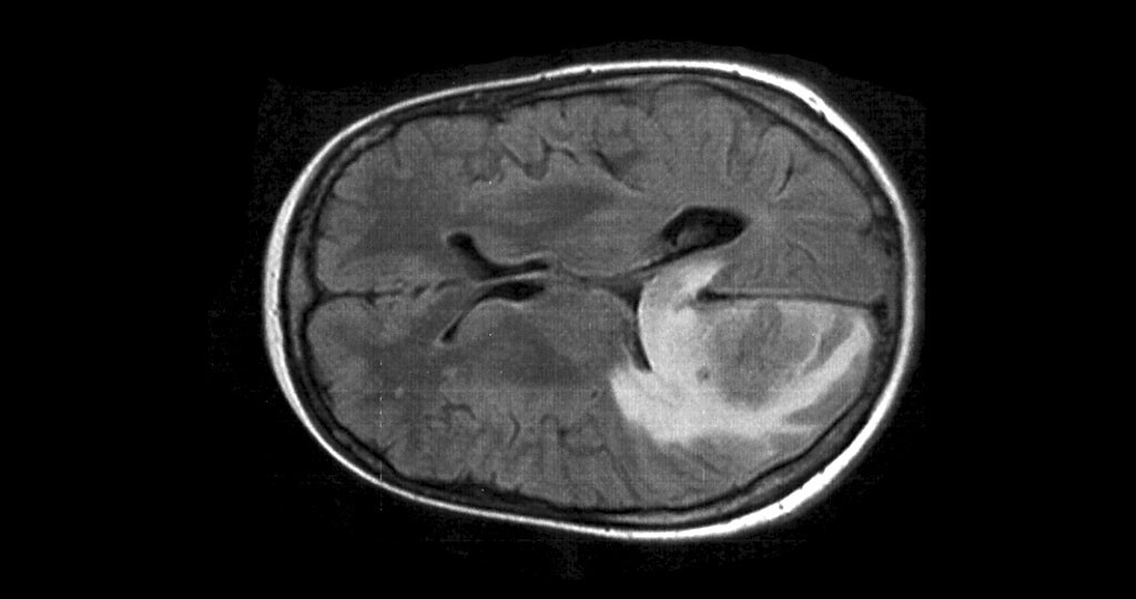
Bestrahlung Gegen Hirnmetastasen Bei Lungenkrebs

Virtual Screening Based Discovery And Mechanistic Characterization Of The Acylthiourea Mrt 10 Family As Smoothened Antagonists Molecular Pharmacology
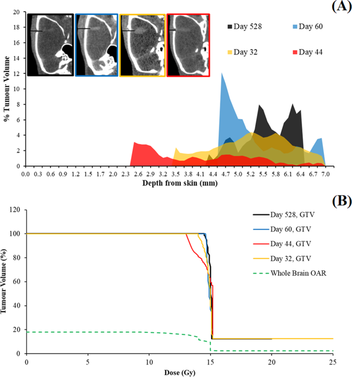
Toward Personalized Synchrotron Microbeam Radiation Therapy Scientific Reports
Q Tbn And9gctaybrvf X7plfz6t6cdp1muvvnfqov9i13p Bxkqs Usqp Cau

File Akustikus Schwannon Rechts Mrt T1km Coronar 001 Jpg Wikimedia Commons
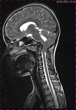
Zufallsbefunde Arnold Chiari Malformation

Volume Measurement Of Liver Metastase Eref Thieme

Magnetic Resonance Imaging Wikipedia

Identification And Analyses Of Extra Cranial And Cranial Rhabdoid Tumor Molecular Subgroups Reveal Tumors With Cytotoxic T Cell Infiltration Sciencedirect
Http Ar Iiarjournals Org Content 40 11 6159 Full Pdf
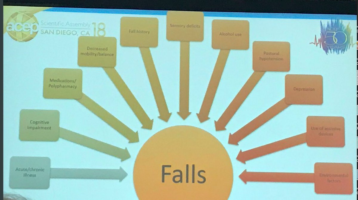
Bernadette Keefe Md Acep18 Via Acep Re Geried Olderpeople Geriatricem Geriem Geriatricer Polypharmacy Too Many Drugs Prescribed Taken Sideeffects Esp Serious In Olderadults Mrt Geriatricednews Clshenvi Opened Geda

Mrt Auditory 01 Vidinio Klausos Kanalo Mrt Mrt Radiologija Radiologija Lt Radiologija
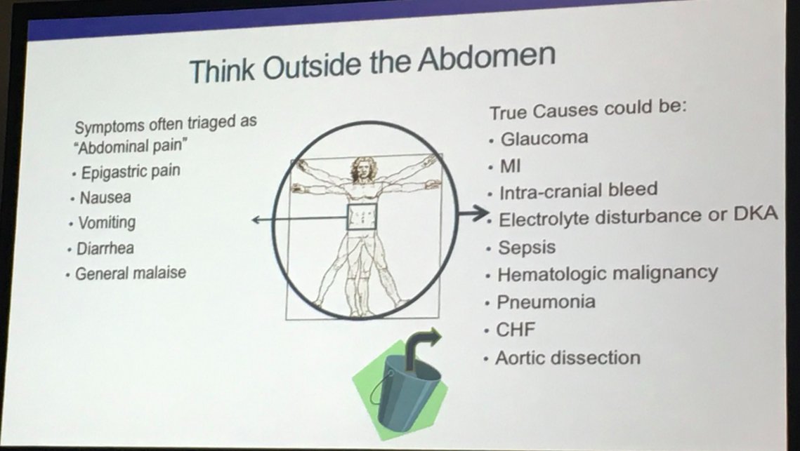
Bernadette Keefe Md Acep18 Acep Geried Geriem Geriatricer Mrt Geriatricednews Clshenvi On Mild Traumatic Brain Injury Mtbi Or Concussion With Or Without Loss Of Consciousness Mild A Misnomer

Clinical Cases Fonar Upright Mri

Gehirn Mrt Atlas Der Menschlichen Anatomie

Mrt Archive Vetradiologie

Cranial Base Surgery Neurochirurgie Luzern Prof Dr Med A Sepehrnia

Ms Diagnose Was Zeigt Eine Mrt Untersuchung An

Gehirn Mrt Atlas Der Menschlichen Anatomie

Bildgebende Verfahren In Der Kopf Hals Diagnostik

Chapter8 1 Mrt 133 Chapter 8 Self Assessment 1 The Nervous System Consists Of The Central Nervous System Cns And The Peripheral Nervous System Pns Course Hero

A D E Ect Of Chemotherapeutic Drugs On 3 H Thymidine Incorporation In Download Scientific Diagram

Nuklearmedizin Braunschweig Celler Strasse 30 Braunschweig Mrt Des Kopfes
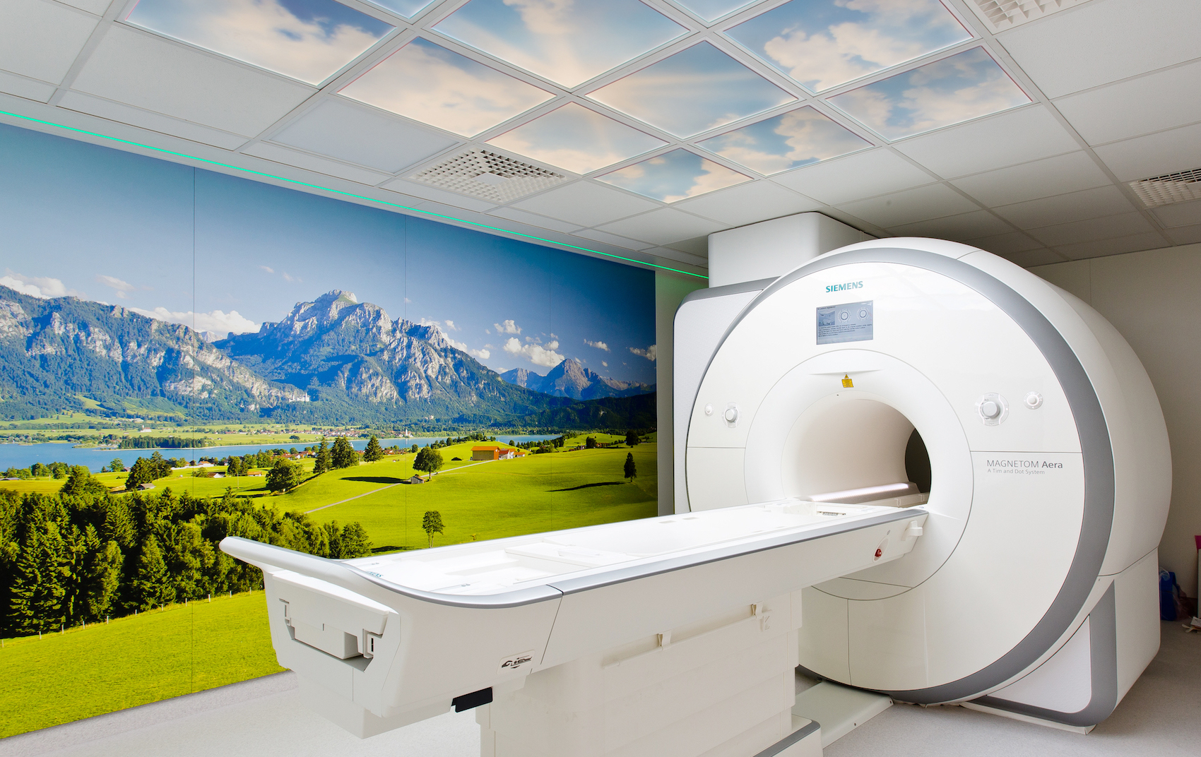
التصوير بالرنين المغناطيسي Mrt Radiologie Baden Baden

Fmy3xk9ck8aunm
Q Tbn And9gcsstgjmu7fsc7q Wkr4jfspojomjhzr6jqaxsk Scpfeto1euef Usqp Cau

Magnetresonanztomographie Mrt Des Kopfes Ct Mrt Institut Berlin

Mrt Untersuchung Alkohol Und Tabak Lassen Das Gehirn Schneller Altern Pz Pharmazeutische Zeitung

Klinische Neuroanatomie Kranielle Mrt Und Ct Atlas Der Magnetresonanztomographie Und Computertomographie Lanfermann Heinrich Raab Peter Kretschmann Hans Joachim Weinrich Wolfgang 本 通販 Amazon

Abb 3 Mrt Eines Fruhgeborenen 31 1 Ssw Am Et T1 Gewichtete Download Scientific Diagram

Magnetic Resonance Tomography Mrt Of Intracranial Tumours Initial Experience With The Use Of The Contrast Medium Gadolinium Dtpa Semantic Scholar
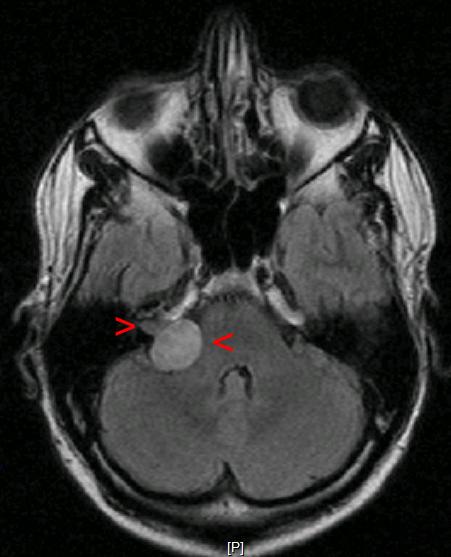
File Akustikusneurinom Mrt Jpg Wikipedia

Nuklearmedizin Braunschweig Celler Strasse 30 Braunschweig Mrt Des Kopfes

Mrt Ct Potsdam Medneo Com Wissenswertes Zu Der Mrt Untersuchung Bei Medneo Gefasse Kopfgefasse

Magnetresonanztomographie Mrt Des Kopfes Ct Mrt Institut Berlin
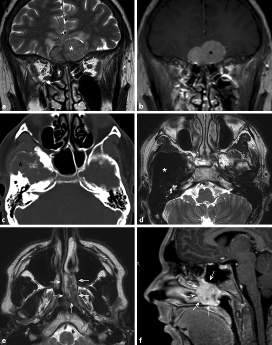
Bildgebung Der Nasennebenhohlen Und Der Frontobasis Springerlink
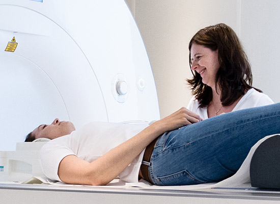
Kernspintomographie Mrt Radiologie Wiesloch Sinsheim
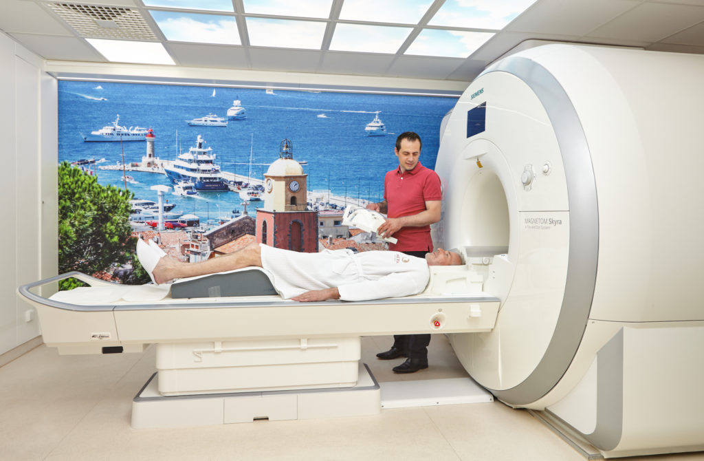
Mrt Kernspintomografie Ablauf Dauer Kosten Praktischarzt

Identification And Analyses Of Extra Cranial And Cranial Rhabdoid Tumor Molecular Subgroups Reveal Tumors With Cytotoxic T Cell Infiltration Sciencedirect
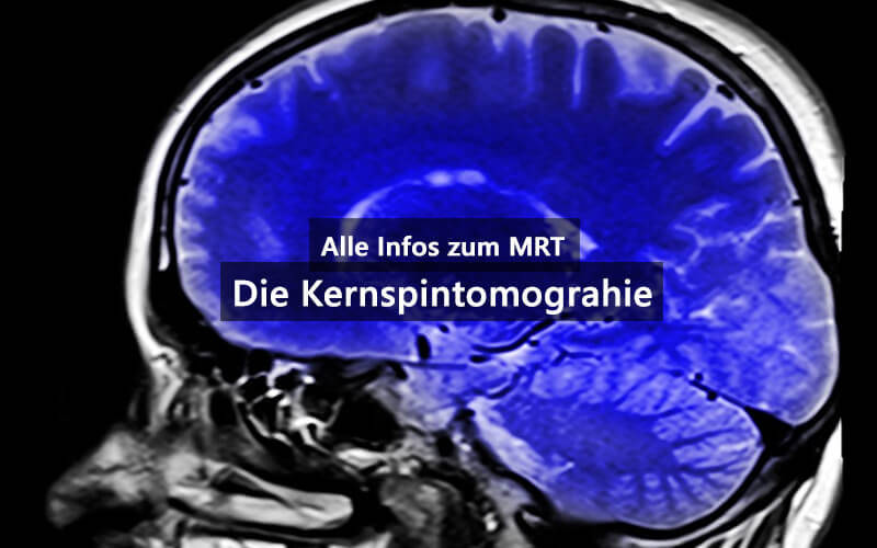
Kopf Mrt Ursachen Fur Kopfschmerzen Schwindel Und Druckgefuhl Finden

Nuklearmedizin Braunschweig Celler Strasse 30 Braunschweig Mrt Des Kopfes
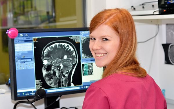
Nuklearmedizin Braunschweig Celler Strasse 30 Braunschweig Mrt Des Kopfes
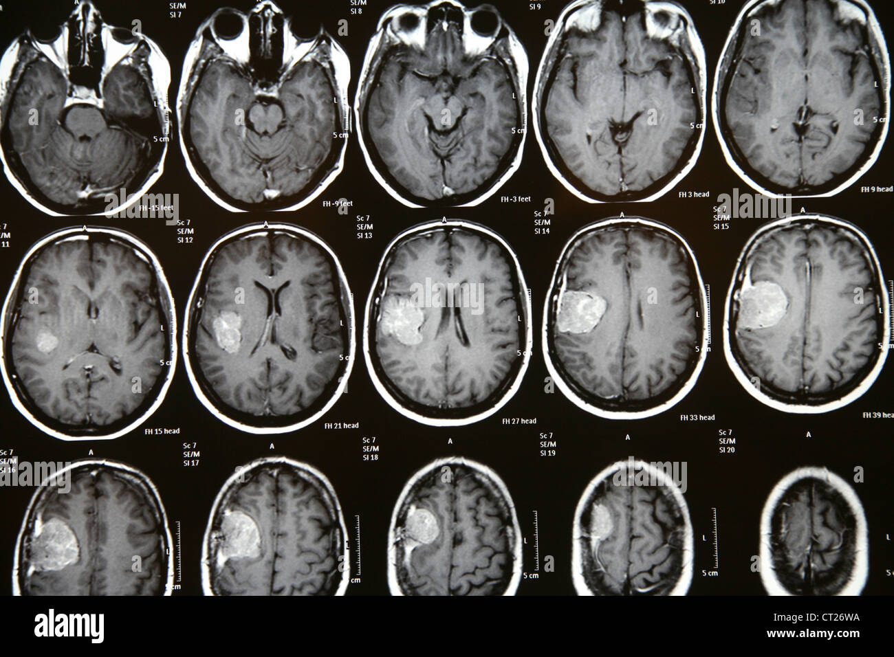
Mri Meningeom Stockfotos Und Bilder Kaufen Alamy
Www Endokrinologie Net Broschueren Php File Files Download Broschueren 2 Hirnanhangdruese Erkrankungen Pdf

Novel Two Mrt Cell Lines Established From Multiple Sites Of A Synchronous Mrt Patient Anticancer Research


