Colony Formation Assay Crystal Violet

An Improved Crystal Violet Assay For Biofilm Quantification In 96 Well Microtitre Plate Biorxiv

Targeted Radiotherapy Potentiates The Cytotoxicity Of A Novel Anti Human Dr5 Monoclonal Antibody And The Adenovirus Encoding Soluble Trail In Prostate Cancer Sciencedirect
Bklotho Overexpression Inhibited Hepatoma Cell Proliferation A Download Scientific Diagram

Overexpression Of Mir 497 Inhibited Colony Formation Ability Of Download Scientific Diagram
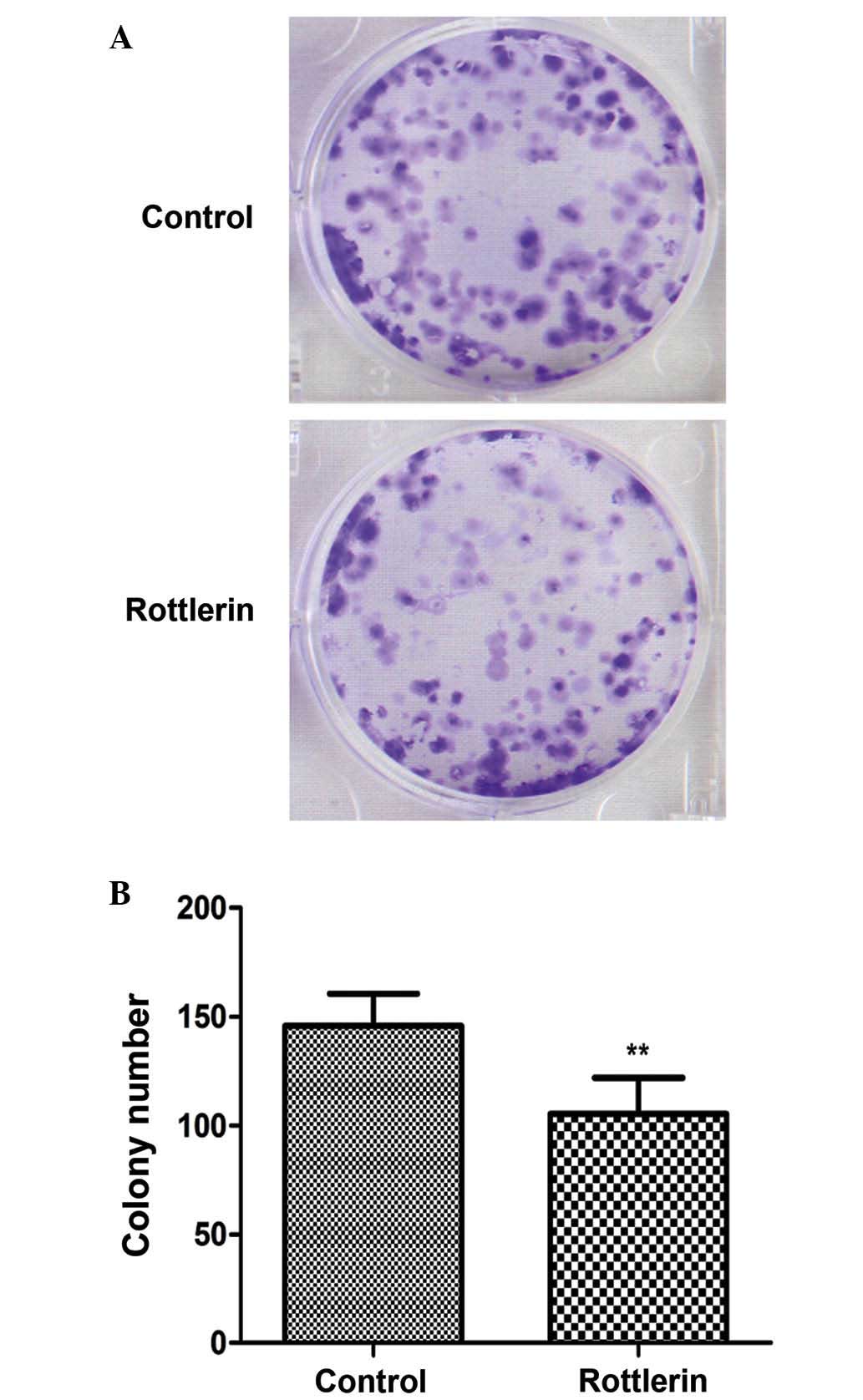
Rottlerin Induced Autophagy Leads To Apoptosis In Bladder Cancer Cells

Facs Mtt And Colony Formation Assays Of Nih3t3 Flagova66 And Nih3t3 Mock Cells
Remove crystal violet carefully and immerse the dishes/plates in tap water to rinse off crystal violet Airdry the dishes/plates on a table cloth at RT for up to a few days Data analysisCount.

Colony formation assay crystal violet. Abstract Colorectal cancer (CRC) is one of the most lethal cancers worldwide The expression of βarrestin2 (βArr2, ARRB2) in CRC has been well investigated;. Abstract Osteosarcoma (OS) is the most common type of primary malignant tumors that originate in the bone Resistance to chemotherapy confers a poor prognosis. Solutions 07% (w/v) Agarose (DNA grade) 1% (w/v) Agar (DNA grade) 0005% Crystal Violet 2X Media % (v/v) Fetal Calf Serum (FCS) BioReagents and Chemicals Fetal Calf Serum (FCS) Crystal Violet Agarose 7 Agar RPMI (or other suitable media) Suggested amounts for soft agar colony formation assay.
Remove the methanol and rinse the cells with H 2 O Add sufficient crystal violet staining solution to cover the cells Incubate the dish for 5 min at room temperature Incubate the dish for 5 min at room temperature. Add 1 ml of PBS containing 4% formaldehyde and 0005% crystal violet to each well Optional Filter the staining solution before applying, otherwise small crystal particles can result in colony. We normally stain colonies with crystal violet, which works well when cells are at a low density and colonies clearly stand out, but at the high plating density needed for this assay I can see that.
A Stain cells with 05 mL Crystal Violet methanol for >1 hour b Wash Crystal violet off by adding 1‐2 mL water to soft agar 4‐5 times c Image on a dissecting microscope d Count colonies > 500 μm or other appropriate diameter Easiest to use. Crystal violet Soft agar colony formation assay University of Virginia True to its name, the simple stain is a very simple staining procedure involving single solution of stain Any basic dye such as methylene blue, safranin, or crystal violet can be used to color the bacterial cells. A Stain cells with 05 mL Crystal Violet methanol for >1 hour b Wash Crystal violet off by adding 1‐2 mL water to soft agar 4‐5 times c Image on a dissecting microscope d Count colonies > 500 μm or other appropriate diameter Easiest to use.
For the colony formation assay, cells (5×10⁵ cells per well) were seeded in 6well plates and transfected with pcDNASEMA3F (2 μg per well) or the control for 24 h The medium was refreshed every 3 days After 2–3 weeks of culture, the colonies were fixed with methanol, stained with 125% crystal violet and counted under a light microscope. Crystal violet solution was used to stain Hela cells colonies after 15 days of treatment with DMSO and inhibitory drug Experimental Design and Results Summary Application. Crystal Violet Staining (for Focus Formation Assay) Place plates on ice Wash two times with icecold 1X PBS Fix cells with icecold methanol (stored at – o C) for 10 minutes Aspirate methanol from plates, move off ice and add enough 05% crystal violet solution (made in 25% methanol and stored at room temperature) to cover bottom of plate.
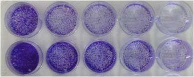
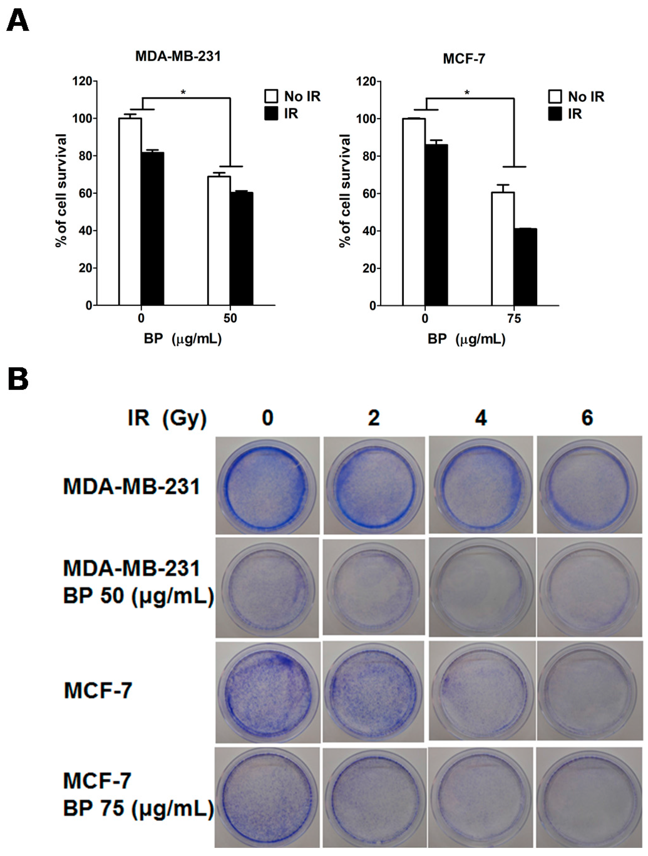
Colonyforming assay 1 Variable Treat cells with cytotoxic agent 5–10 min 10–15 min 2 Observe cells using brightfield microscope 3 Harvest cells 4 30 min Count cells and plate 0 cells/ well 5 Incubate at 37°C 1–2 wk 6 min Fix colonies with 100% methanol 7 Incubate cells in crystal violet 5 min 8 Rinse in water 2 min 9 Invert onto tissue to dry Overnight. Colony forming or clonogenic assay is an in vitro quantitative technique to examine the capability of a single cell to grow into a large colony through clonal expansion Clonogenic activity is a sensitive indicator of undifferentiated cancer stem cells Here, we described the colony forming ability of the isolated breast cancer stem cells from the total population of cancer cells using doublelayered, soft agarosebased assay. Crystal violet cell proliferation assay After irradiation, 2500 cells in one ml growth medium were seeded into 24well plates in quadruplicate for each dose Cells were incubated at 37 °C and 5% CO 2 for 5 days This was the time point at which the cells in the shamirradiated plates nearly reached confluency.
The Crystal Violet assay assumes that all cells that are attached to the plate are "alive" and that all cells that detach are "dead" Crystal violet is a triarylmethane dye that binds to ribose type molecules such as DNA in nuclei Crystal Violet assay protocol summary remove culture medium wash cells add Crystal Violet staining solution incubate for mins wash cells and air dry plate. Most of the nonresistant cells are dead after 34 days of puromycin selection However, it takes 23 weeks for the drugresistant cells to form a visible colony The assay should be stopped when the colonies are clearly visible even without looking under the microscope Stain the colonies with crystal violet and count them if so desired Notes. For colony formation assay, singlecell suspensions were seeded in 6 well plate (NEST Biotechnology Co LTD) at a density of 500 cells/well with 10% FBS containedmedium After 7 days incubation, the colonies were fixed with 4% paraformaldehyde and stained by crystal violet The numbers of colonies were counted Each assay was performed in triplicate.
The colony formation assay is typically carried out in a multiwell format Single cells are seeded in each well at low density in the presence of various concentrations of compounds or vehicle control After a set growth period, colonies are fixed and stained with Crystal Violet, then the number of colonies is scored by visual inspection. Crystal violet assay is a quick and reliable screening method that is suitable for the examination of the impact of chemotherapeutics or other compounds on cell survival and growth inhibition 47 8 Genotoxicity A Predictable Risk to Our Actual World. Results According to MTT, crystal violet and colony formation assay results, EVO significantly inhibited the cell proliferation in a dosedependent manner Hoechst staining assay revealed that EVO induced cell apoptosis in a concentrationdependent manner Moreover, EVO inhibited the migration and invasion of the osteosarcoma cells.
This video protocol provides stepbystep instructions on how to consistently perform the Colony Forming Cell (CFC) Assay Tips are provided throughout the v. 0005% Crystal Violet 2X Media % (v/v) Fetal Calf Serum (FCS) BioReagents and Chemicals Fetal Calf Serum (FCS) Agar Crystal Violet Agarose RPMI (or other suitable media) Suggested amounts for soft agar colony formation assay Culture Dish 96 well 48 well 24 well 6 well 35 mm 60 mm 100 mm Base and Top Agar Volume (mL/well). Tumor necrosis factorrelated apoptosisinducing ligand (TRAIL) can induce substantial cytotoxicity in tumor cells but rarely exert cytotoxic activity on nontransformed cells In the present study, we therefore evaluated interactions between TRAIL and IER3 via coimmunoprecipitation and immunofluorescence analyses, leading us to determine that these two proteins were able to drive the.
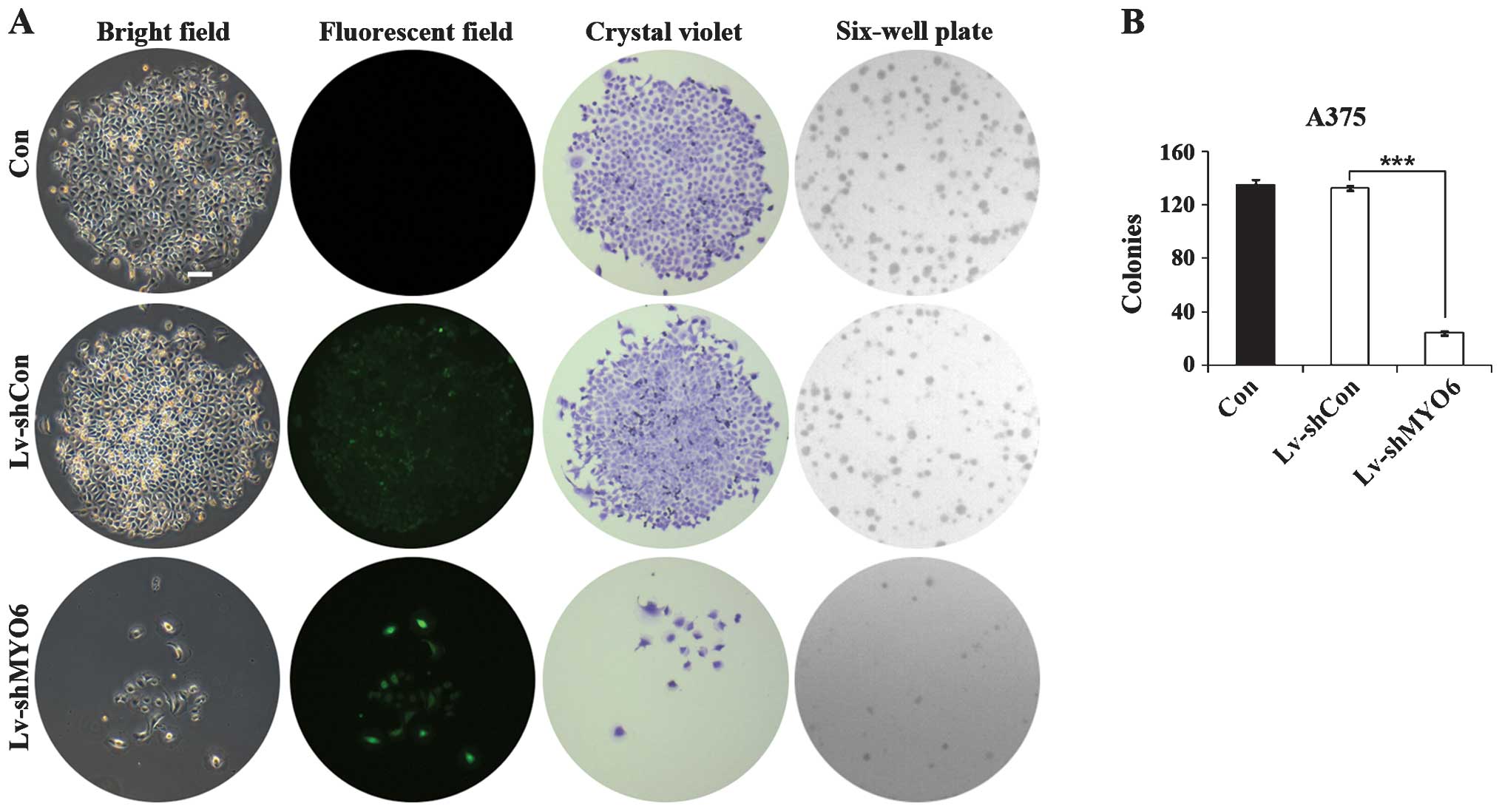
(B) Representative images of colony formation assays of Con, LvshCon and LvshUSP39 infected SMMC7721 cells From top to bottom crystals violet staining, bright field and GFP of single colony, and crystals violet staining of sixwell plate Scale bar, 25 μm (C) Colony numbers of Con, LvshCon and LvshUSP39 infected SMMC7721 cells. Add 05% crystal violet solution and incubate at RT for 2 h Add 10 ml medium with 10% FBS, and detach the cells by pipetting Remove crystal violet carefully and immerse the dishes/plates in tap water to rinse off crystal violet Airdry the dishes/plates on a table cloth at RT for up to a few days Data analysis. Filter 01% crystal violet solution through a 022 μm filter to remove any precipitates Stain each well with 1 mL of 01% crystal violet for 10 min at room temperature Gently remove the stain Wash cells 3x with 1 mL of PBS, being careful not to disturb the colonies Count the colonies for at least 2 of the dilutions.
The cells were fixed, and colony morphologies were scored Colonies were fixed with 70% ethanol and were stained with 05% crystal violet A cell colony was defiend as a groupe formation of at least 50 cells and counted using Image J software (National Institutes of Health, Bethesda, Maryland, USA). Circular RNA circSEC24A Promotes Cutaneous Squamous Cell Carcinoma Progression by Regulating miR1193/MAP3K9 Axis. A review of the Crystal Violet Solution for Colony Formation Assays Unbiased reviews by scientists available at Biocomparecom.
The cells were fixed, and colony morphologies were scored Colonies were fixed with 70% ethanol and were stained with 05% crystal violet A cell colony was defiend as a groupe formation of at least 50 cells and counted using Image J software (National Institutes of Health, Bethesda, Maryland, USA). Methanol from plates, move off ice and add enough 05% crystal violet solution (made in 25% methanol and stored at room temperature) to cover bottom of plate. K Crystal Violet Cell Cytotoxicity Assay Kit FOR RESEARCH USE ONLY!.
We also employed a colony formation assay in soft agar to evaluate changes in anchorage independent growth after loss of CDCP1 In this assay, 44fold more colonies were observed in CDCP1silenced cells compared to controls (Fig 3b) In addition to the increase in colony numbers, colony size was also increased. Colonyforming assay 1 Variable Treat cells with cytotoxic agent 5–10 min 10–15 min 2 Observe cells using brightfield microscope 3 Harvest cells 4 30 min Count cells and plate 0 cells/ well 5 Incubate at 37°C 1–2 wk 6 min Fix colonies with 100% methanol 7 Incubate cells in crystal violet 5 min 8 Rinse in water 2 min 9 Invert onto tissue to dry Overnight. Crystal violet Assay Kit (Cell viability) (ab) Abcam Remove the methanol and rinse the cells with H2O Add sufficient crystal violet staining solution to cover the cells Incubate the dish for 5 min at room temperature 8 Wash the cells with H2O until excess dye is removed Measuring Survival of Adherent Cells with the Colony.
Crystal violet Assay Kit (Cell viability) (ab) Abcam Remove the methanol and rinse the cells with H2O Add sufficient crystal violet staining solution to cover the cells Incubate the dish for 5 min at room temperature 8 Wash the cells with H2O until excess dye is removed Measuring Survival of Adherent Cells with the Colony. 1000 assays, Store kit at °C) I Introduction Crystal violet (CV) cell cytotoxicity assay is one of the common methods used to detect cell viability or drug cytotoxicity CV is a triarylmethane dye that can bind to ribose type molecules such as DNA in nuclei. Chronic Pseudomonas aeruginosa lung infections in cystic fibrosis (CF) evolve to generate environmentally adapted biofilm communities, leading to increased patient morbidity and mortality OligoG CF5/, a lowmolecularweight inhaled alginate oligomer therapy, is currently in phase IIb/III clinical trials in CF patients Experimental evolution of P aeruginosa in response to OligoG CF5/.
155 S Milpitas Blvd, Milpitas, CA USA T (408) F (408) wwwbiovisioncom tech@biovisioncom Crystal Violet Cell Cytotoxicity Assay Kit. Molar Mass g·mol−1. Staining colonies with crystal violet 1 Count colonies on each gel before staining Count twice Divide plates into quarters to help with high counts 2 Add 03 ml of 01% crystal violet solution to each plate 01% Crystal Violet solution = 3 mL 05% crystal violet 15 mL ethanol 105 mL water 3 Incubate 15 min on a level surface Rotate gels.
Dye Color When dissolved in water, the dye has a blueviolet color Ingredient For Equine Herbal Products To Treat Digestion, Performance, Respiratory Issues And Thrush;. After fixation, 10min cell staining was implemented with 01% crystal violet (G1064, Solarbio) The number of colonies with more than 50 cells was counted under a microscope (Leica, Germany) e Colony formation assay detecting the colony formation of A549 and H460 cells treated with EVs derived from H1299 or H522 cells transfected with miR. Only a fraction of seeded cells retains the capacity to produce colonies Before or after treatment, cells are seeded out in appropriate dilutions to form colonies in 13 weeks Colonies are fixed with glutaraldehyde (60% v/v), stained with crystal violet (05% w/v) and counted using a stereomicroscope.
A colony formation assay using crystal violet staining was performed to compare cell proliferation, while woundhealing and Transwell assays were performed to compare cell migration and invasion Subsequently, bioinformatics and a luciferase reporter gene assay were used to investigate the effect of miR23a on sine oculis homeobox homolog 1 (SIX1) expression, and the biological function of SIX1 was analyzed. Colony formation as assessed by traditional manual counting was compared with the results from the crystal violet dissolution assay The overall bivariate correlation of the data was determined (r = 0743, p < 001). Quantification of colony formation measuring absorption Colony formation was quantified following the method described by Kueng and coworkers In brief, the crystal violet staining of cells from each well was solubilized using 1 ml of 10% acetic acid and the absorbance (optical density) of the solution was measured on a Synergy H1 hybrid fluorescence platereader (BioTek, Winooski, VT, USA) at a wavelength of 590 nm.
Plates were then stained with crystal violet, and colonies consisting of 50 or more cells were manually counted Results were normalized to the colonyforming efficiency of the vehicle control The survival fraction was calculated based on the number of colonies formed in drugtreated cells relative to that of the untreated control. Vandersickel, Veerle, Jacobus Slabbert, Hubert Thierens, and Anne Vral 11 “Comparison of the Colony Formation and Crystal Violet Cell Proliferation Assays to Determine Cellular Radiosensitivity in a Repairdeficient MCF10A Cell Line” Radiation Measurements 46 (1) 72–75. This method demonstrates that cancer stem cells can survive and generate colony growth in an anchorageindependent culture model The 0005% crystal violet solution is used in this assay to visualize the generated colonies.
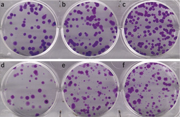
Crystal Violet Cell Colony Staining Crystal Violet Cell Colony Staining 1L Fixing/Staining solution 05 g Crystal Violet (005% w/v) 27 ml 37% Formaldehyde (1%) 100 mL 10X PBS (1X) 10 mL Methanol (1%) 863 dH to 1L 1) Remove media (do not wash cells) 2) Add staining solution to cover dish 3) Stain for min at room temperature 4) Remove fix/stain solution and save 5) Wash dishes one at a time by dipping into bucket of water in the sink with the water continuing to run 6) Air dry. C and d Colony formation assay of Ishikawa (c) and HEC1A (d) cells transfected with NT siRNA or GPR64 siRNA Samples from each treatment were transferred to flatbottomed 24well plates and incubated Cells were fixed and stained with crystal violet The average colony formation number was quantified with crystal violet stained cells. A clonogenic assay, also known as a colony formation assay is an in vitro cell survival assay It assesses the ability of single cells to survive and reproduce to form colonies 1 This assay was first described in the 1950s, where it was used to study the effects of radiation on cancer cell survival and growth and has subsequently played an essential role in radiobiology 2.
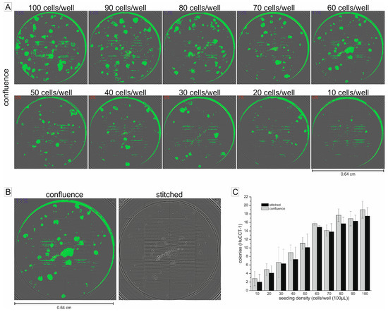
Crystal Violet Assay Kit ab is used for cytoxicity and cell viability studies with adherent cell cultures The Crystal Violet assay is based on staining cells that are attached to cell culture plates It relies on the detachment of adherent cells from cell culture plates during cell death During the assay, dead detached cells are washed. SRB assay is also used to evaluate colony formation and colony extinction Advantages Crystal violet assay is a quick and reliable screening method that is suitable for the examination of the impact of chemotherapeutics or other compounds on cell survival and growth inhibition. Conducting a colony formation assay in a 96well microplate using upright brightfield microscopy The fluorescent properties of Crystal Violet are first utilized to define the colony area, while Hoechst is used to quantify the number of cells within the colony Quantitative microscopy using the Cytation 7 Cell Imaging MultiMode.
Colony formation assay For the colony formation assay, a total of 500 cells/well were seeded into 6well plates and incubated for 15 days Cell colonies were fixed with methanol, then the colonies were stained with 05% crystal violet (Sigma, Darmstadt, Germany) for 30 minutes at room temperature The total number of colonies were photographed and counted Transwell assay. Figure 1 A graphic flowchart of the colonyforming assay for adherent cells. Solutions 07% (w/v) Agarose (DNA grade) 1% (w/v) Agar (DNA grade) 0005% Crystal Violet 2X Media % (v/v) Fetal Calf Serum (FCS) BioReagents and Chemicals Fetal Calf Serum (FCS) Crystal Violet Agarose 7 Agar RPMI (or other suitable media) Suggested amounts for soft agar colony formation assay.
The Crystal Violet assay assumes that all cells that are attached to the plate are "alive" and that all cells that detach are "dead" Crystal violet is a triarylmethane dye that binds to ribose type molecules such as DNA in nuclei Crystal Violet assay protocol summary remove culture medium wash cells add Crystal Violet staining solution incubate for mins wash cells and air dry plate. Principles And Purpose Crystal Violet Assay for Determining Viability of Cultured Cells;. 25 Colony formation assay Cells were treated in the presence of irradiation Fourteen days later, the cells were stained with 05% crystal violet in absolute ethanol, and colonies that contained more than 50 cells were counted under an inverted microscope (Olympus, Tokyo, Japan) 26 Cell apoptosis analysis.
The assay should be stopped when the colonies are clearly visible even without looking under the microscope Stain the colonies with crystal violet and count them if so desired.
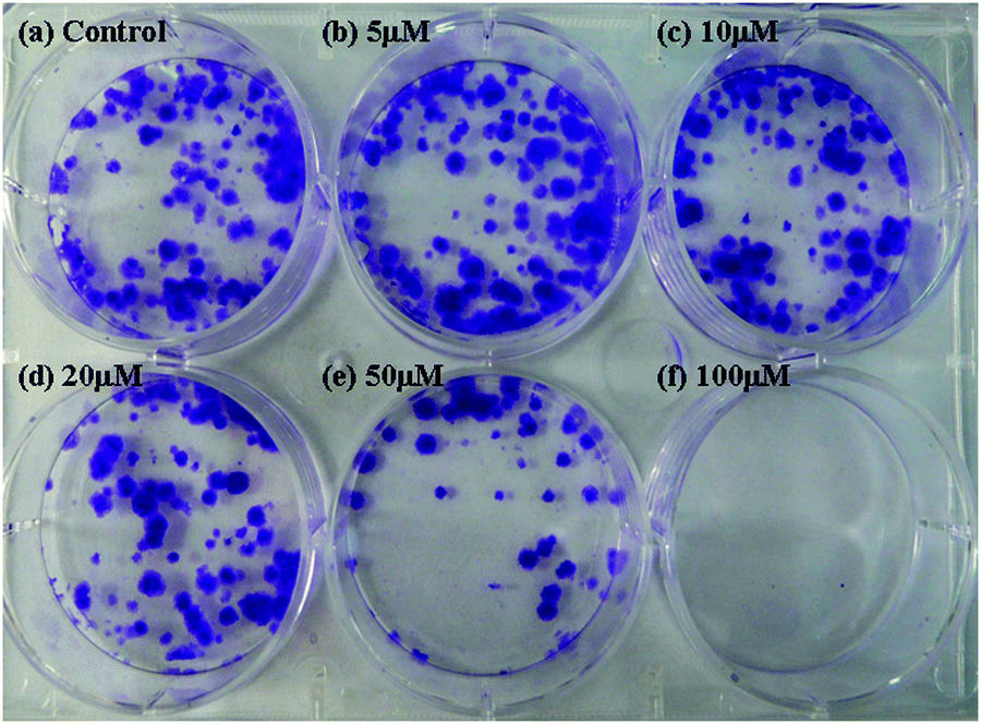
Two Dpa Based Zinc Ii Complexes As Potential Anticancer Agents Nuclease Activity Cytotoxicity And Apoptosis Studies New Journal Of Chemistry Rsc Publishing

Foxm1 Mediates Resistance To Herceptin And Paclitaxel Cancer Research
Onlinelibrary Wiley Com Doi Pdf 10 2164 Jandrol 109
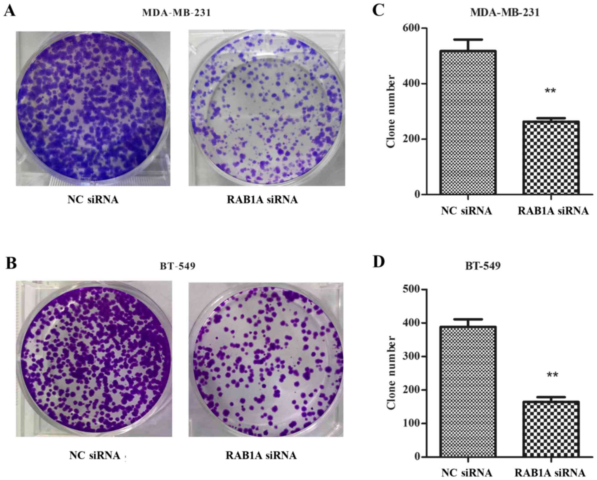
Inhibition Of Rab1a Suppresses Epithelial Mesenchymal Transition And Proliferation Of Triple Negative Breast Cancer Cells
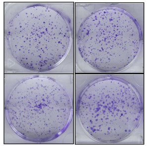
Clonogenic Assay Creative Bioarray Creative Bioarray

Addgene Colony Formation Titering Assay

Addgene Colony Formation Titering Assay
Plos One Targeting Insulin Like Growth Factor 1 Receptor Inhibits Pancreatic Cancer Growth And Metastasis
Hedgehog Signaling Pathway Regulates Human Pancreatic Cancer Cell Proliferation And Metastasis

Figure 3 From Activities Of A Novel Schiff Base Copper Ii Complex On Growth Inhibition And Apoptosis Induction Toward Mcf 7 Human Breast Cancer Cells Via Mitochondrial Pathway Semantic Scholar
Ijms Free Full Text Miniaturization Of The Clonogenic Assay Using Confluence Measurement Html

Knockdown Of Tctn1 Inhibits Colony Formation Of The Pc 3 And Du145 Download Scientific Diagram

Cytosmart Clonogenic Assay What Why And How

Colony Formation Assay Cells Were Seeded And Incubated In The Download Scientific Diagram

Colony Formation Assay Of Holoclones Parent Pc3 And Paraclones Four Download Scientific Diagram

A Colony Forming Assay The Cells Were Plated At A Density Of 100 Download Scientific Diagram
Plos One Artonin E Induces Apoptosis Via Mitochondrial Dysregulation In Skov 3 Ovarian Cancer Cells

Colony Formation Assay Of Bmscs And Muscs
Colony Forming Assay A Colony Forming Assay Of Primary Hcecs After Download Scientific Diagram
How To Do Cancer Cell U87 Cell Colony Formation Assay
Mir 34a And Mir 3 Inhibit Survivin Expression To Control Cell Proliferation And Survival In Human Osteosarcoma Cells
Ijms Free Full Text Graphene Oxide Silver Nanocomposite Enhances Cytotoxic And Apoptotic Potential Of Salinomycin In Human Ovarian Cancer Stem Cells Ovcscs A Novel Approach For Cancer Therapy Html

Comparison Of The Colony Formation And Crystal Violet Cell Proliferation Assays To Determine Cellular Radiosensitivity In A Repair Deficient Mcf10a Cell Line Sciencedirect
Www Biotek Com Assets Tech Resources High Throuput fluorescent colony formation App Note Final Pdf

Cytosmart Clonogenic Assay What Why And How

Colony Formation And Invasion Assay Of Hctshdapk And Hctwtdapk Cells Download Scientific Diagram

Full Text Da0324 An Inhibitor Of Nuclear Factor Kb Activation Demonstrat Dddt

Clonogenic Assay After Different Treatments Of Panc1 Tumor Cells Download Scientific Diagram
Q Tbn And9gcqxskutgvgj0wtkdb9ltsyc04dfyokvhremigd6fsto75eai Li Usqp Cau
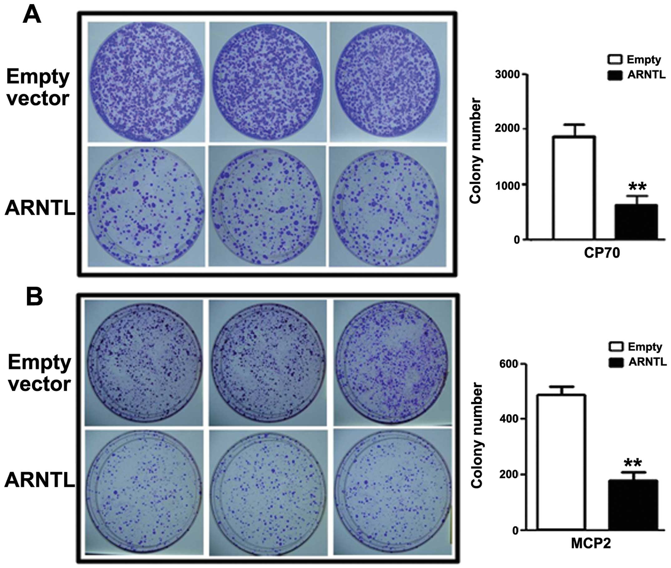
Epigenetic Silencing Of Arntl A Circadian Gene And Potential Tumor Suppressor In Ovarian Cancer
5 Methoxyhydnocarpin Shows Selective Anti Cancer Effects And Induces Apoptosis In Thp 1 Human Leukemia Cancer Cells Via Mitochondrial Disruption Suppression Of Cell Migration And Invasion And Cell Cycle Arrest Bangladesh Journal Of

Figure 15 From Mixed Ligand Copper Ii Schiff Base Complexes The Role Of The Co Ligand In Dna Binding Dna Cleavage Protein Binding And Cytotoxicity Semantic Scholar

Lentivirus Mediated Silencing Of Ubiquitin Specific Peptidase 39 Inhibits Cell Proliferation Of Human Hepatocellular Carcinoma Cells In Vitro

Clonogenic Assay A B16 F10 B 058 C Jr8 Cells Were Treated Download Scientific Diagram

Full Text Progranulin Modulates Cholangiocarcinoma Cell Proliferation Apoptosis Ott
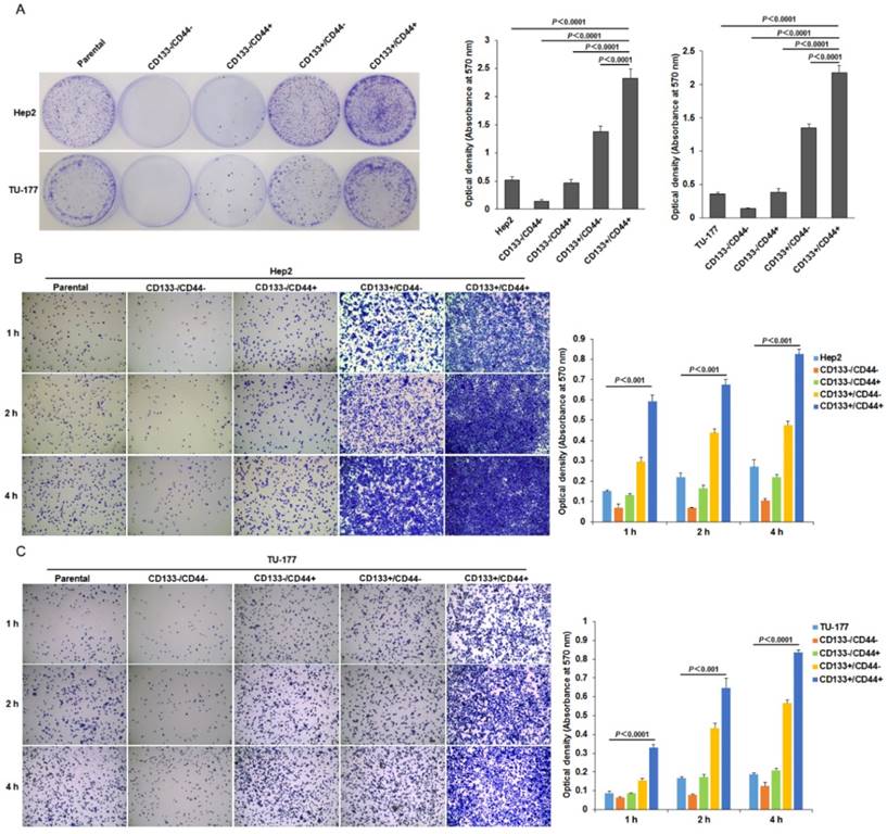
Identification And Characterization Of Cd133 Cd44 Cancer Stem Cells From Human Laryngeal Squamous Cell Carcinoma Cell Lines

Clonogenic Assay Creative Bioarray Creative Bioarray
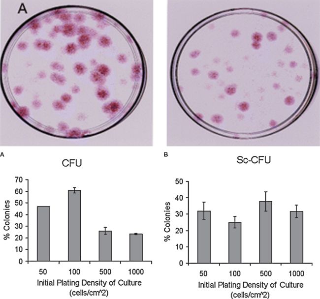
Colony Forming Unit Assays For Mscs Springerlink
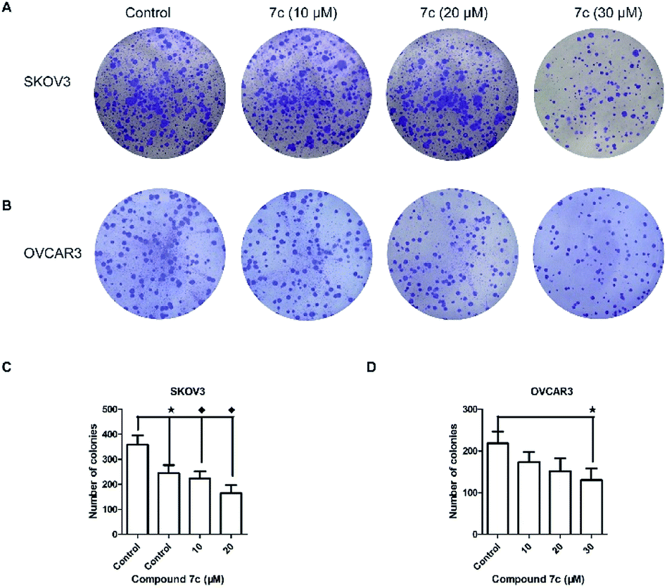
Synthesis And Discovery Of 18b Glycyrrhetinic Acid Derivatives Inhibiting Cancer Stem Cell Properties In Ovarian Cancer Cells Rsc Advances Rsc Publishing Doi 10 1039 C9rad

Xmlinkhub
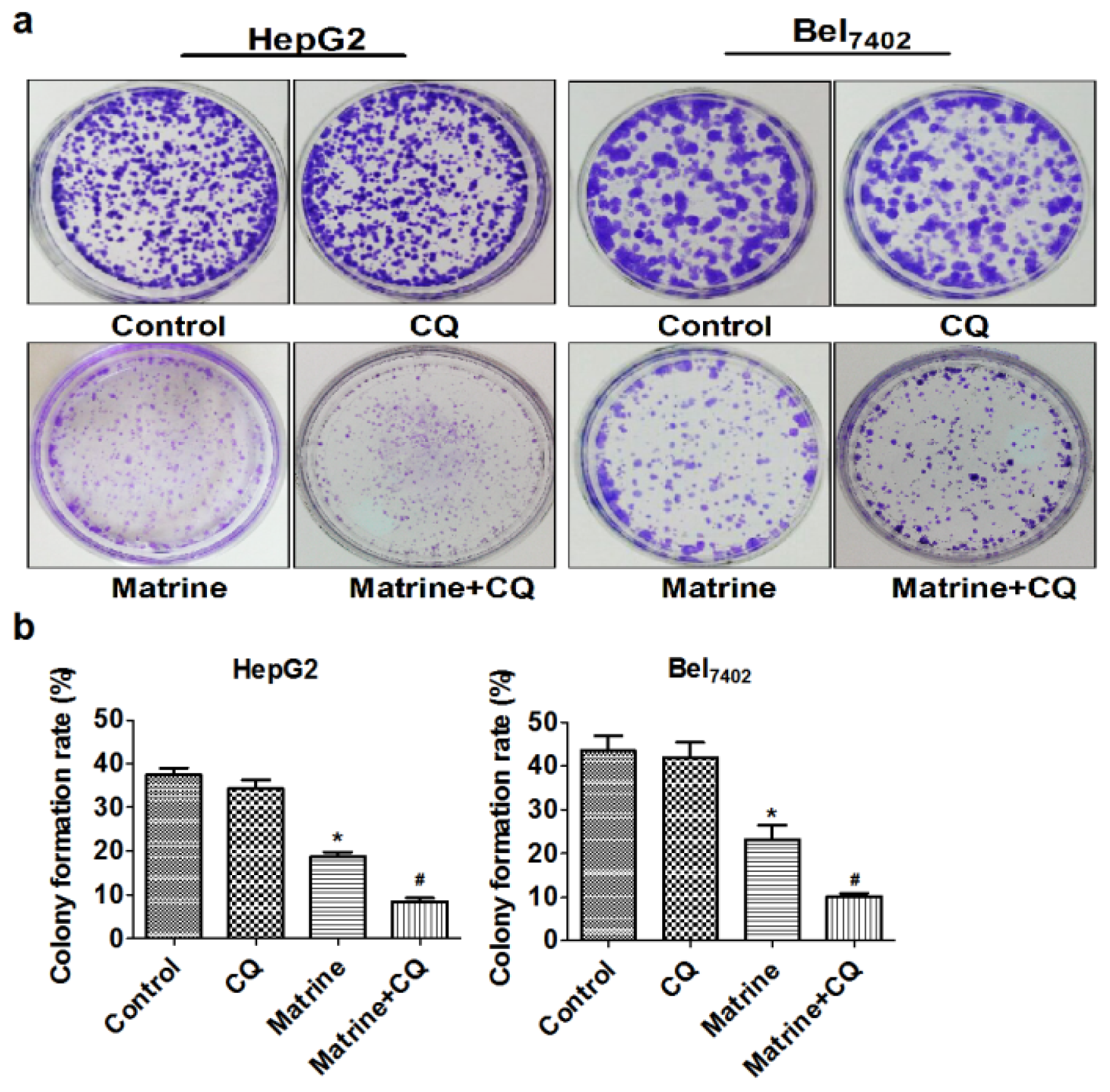
Ijms Free Full Text Blocking Autophagic Flux Enhances Matrine Induced Apoptosis In Human Hepatoma Cells Html
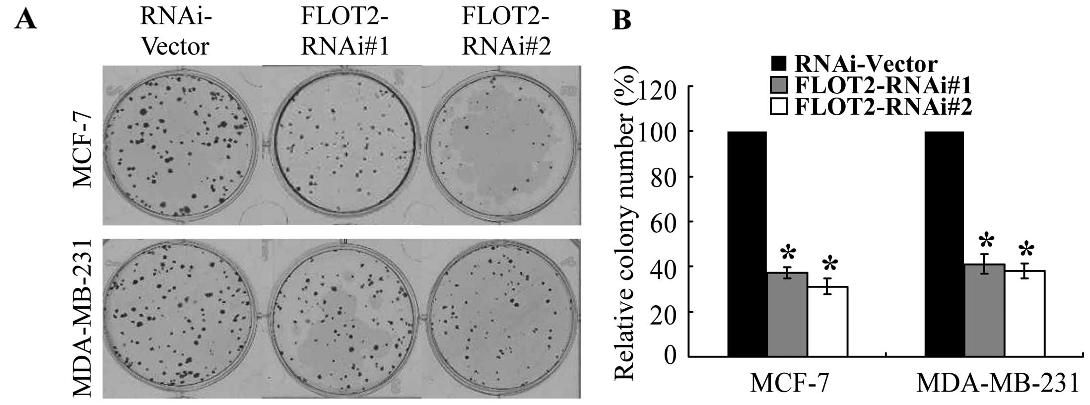
Knockdown Of Flotillin 2 Impairs The Proliferation Of Breast Cancer Cells Through Modulation Of Akt Foxo Signaling
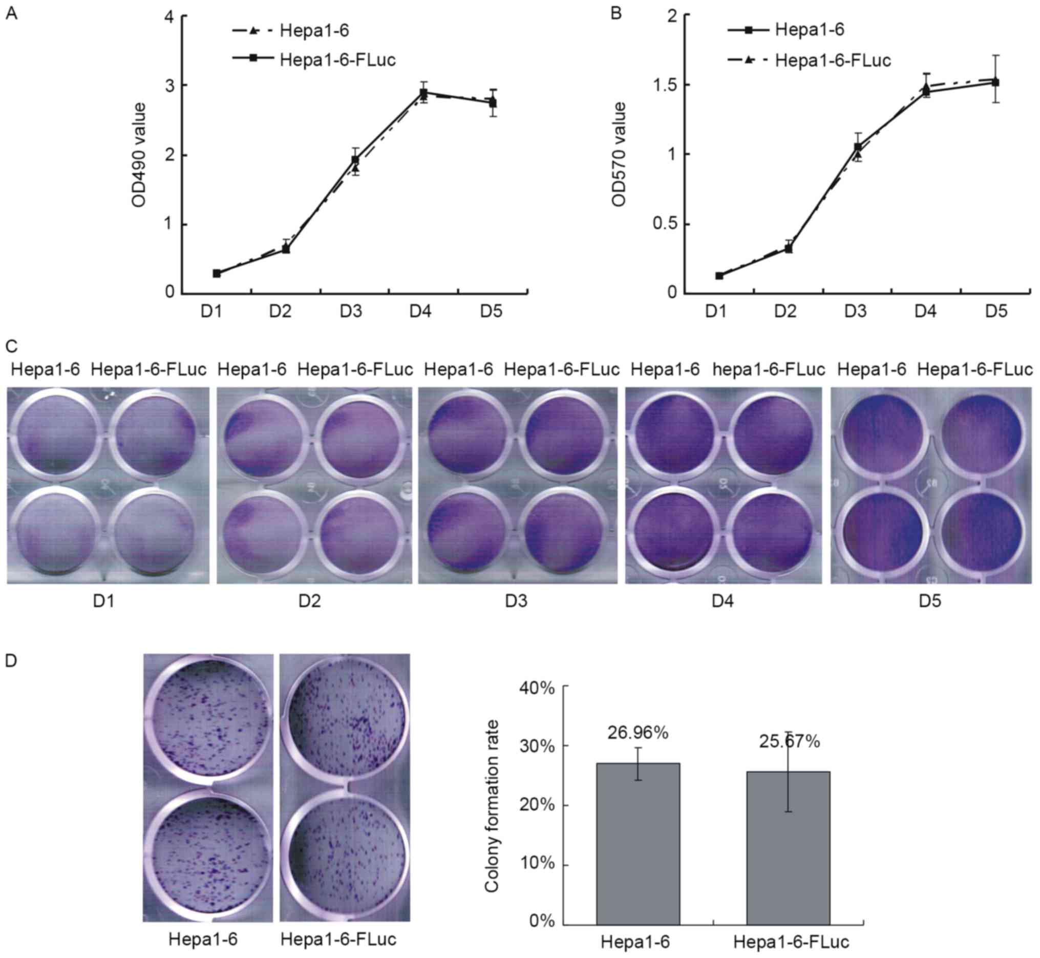
Hepa1 6 Fluc Cell Line With The Stable Expression Of Firefly Luciferase Retains Its Primary Properties With Promising Bioluminescence Imaging Ability
Plos One Secreted Frizzled Related Protein 4 Inhibits Glioma Stem Like Cells By Reversing Epithelial To Mesenchymal Transition Inducing Apoptosis And Decreasing Cancer Stem Cell Properties
Artscimedia Case Edu Wp Content Uploads Sites 198 16 10 Soft Agar Assay Protocol Pdf
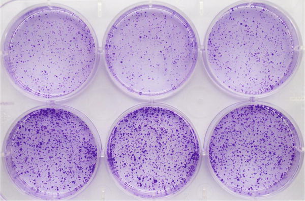
Analysis Of Epithelial Mesenchymal Transition Induced By Overexpression Of Twist Springerlink

Bas 4 Reduced C6 Cell Colony Formation As Analyzed By A Colony Forming Download Scientific Diagram
Www Mdpi Com 73 4409 8 2 117 Pdf

Clonogenic Assay For Dnmt3l Overexpressing Hela Cells A Download Scientific Diagram
Clonogenic Assay Of Cells In Vitro Nature Protocols

Cell Type Dependent Function Of Lats1 2 In Cancer Cell Growth Abstract Europe Pmc

A The Colony Formation Assay Was Visualized And Evaluated By Crystal Download Scientific Diagram

Clonogenic Assay Of Sacc Cells Transfected With Pim 1 And Control Sirna Download Scientific Diagram

High Throughput Fluorescent Colony Formation Assay January 10
Q Tbn And9gctmvp9fr7qyyqokcnx9gi48bab1whgbinevei1czx Lrcii0wqm Usqp Cau
Selective Cytotoxic Effect Of Plantago Lanceolata L Against Breast Cancer Cells Journal Of The Egyptian National Cancer Institute Full Text
Knockdown Of Myosin Vi By Lentivirus Mediated Short Hairpin Rna Suppresses Proliferation Of Melanoma
Plos One The Histone Demethylase Jarid1b Kdm5b Is A Novel Component Of The Rb Pathway And Associates With E2f Target Genes In Mefs During Senescence

High Throughput Fluorescent Colony Formation Assay Metrolab Blog

Full Text Downregulation Of Deptor Inhibits The Proliferation Migration And Su Ott
Ijms Free Full Text Miniaturization Of The Clonogenic Assay Using Confluence Measurement Html

Tuft1 Promotes Osteosarcoma Cell Proliferation And Predicts Poor Prognosis In Osteosarcoma Patients In Open Life Sciences Volume 13 Issue 1 18
Plos One G9a Inhibition Induces Autophagic Cell Death Via Ampk Mtor Pathway In Bladder Transitional Cell Carcinoma

Growth Of Lacscs And Hif1a Kd Induced Cells As Measured By Colony Download Scientific Diagram

Oridonin Inhibited Colony Formation In Glioma Cells A Crystal Violet Download Scientific Diagram

Proliferation Capacity A Macroscopic And Microscopic View Of Download Scientific Diagram

Clonogenic Assay An Overview Sciencedirect Topics

Biofilm Formation Assay In Pseudomonas Syringae

Full Text Inhibition Of Foxo1 By Small Interfering Rna Enhances Proliferation An Ott

An Improved Crystal Violet Assay For Biofilm Quantification In 96 Well Microtitre Plate Biorxiv

High Throughput Fluorescent Colony Formation Assay Metrolab Blog

High Throughput Fluorescent Colony Formation Assay January 10

Clonogenic Assay What Why And How Cytosmart

Comparison Of The Colony Formation And Crystal Violet Cell Proliferation Assays To Determine Cellular Radiosensitivity In A Repair Deficient Mcf10a Cell Line Sciencedirect

Inhibitor Of The Spindle Assembly Checkpoint Surpasses Apoptosis Sensitizer In Synergy With Taxanes Biorxiv

Overexpression Of Pik3r1 Promotes Hepatocellular Carcinoma Progression

Colony Formation And Wound Healing Assay A Colony Formation Assay Download Scientific Diagram

Crystal Violet Solution For Colony Formation Assays Biocompare Com Kit Reagent Review

Full Text Paeoniflorin Suppresses Pancreatic Cancer Cell Growth By Upregulating Dddt
Q Tbn And9gctld 0 Ai4aat2aqd6kerhee5w6pnz D8ficdr6fganhel1iuxq Usqp Cau

6 Gingerol Decreases Clonogenicity And Radioresistance Of Human Prostate Cancer Cells Clinical Oncology And Research Science Repository Open Access
Clonogenic Assay Of Cells In Vitro Colony Formation Assay 네이버 블로그
Antitumor Activity Of Az64 Via G2 M Arrest In Non Small Cell Lung Cancer
Molecules Free Full Text Anti Tumor And Radiosensitization Effects Of N Butylidenephthalide On Human Breast Cancer Cells Html

Cytosmart Clonogenic Assay What Why And How

Figure 2 From Small Interfering Rna Sirna Mediated Knockdown Of Notch1 Suppresses Tumor Growth And Enhances The Effect Of Il 2 Immunotherapy In Malignant Melanoma Semantic Scholar
Plos One Curcumin Conjugated With Plga Potentiates Sustainability Anti Proliferative Activity And Apoptosis In Human Colon Carcinoma Cells

Dose Dependent Colony Forming Assay Of Ssbc Different Concentration Of Download Scientific Diagram

Figure 1 From 2 Hydroxyflavanone A Novel Strategy For Targeting Breast Cancer Semantic Scholar
Gli Inhibitor Gant61 Kills Melanoma Cells And Acts In Synergy With Obatoclax

Hypoxia Increases Colony Forming Ability The Colony Forming Ability Of Download Scientific Diagram

Clonogenic Assay Youtube
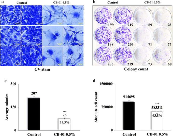
Integration Of Conventional Cell Viability Assays For Reliable And Reproducible Read Outs Experimental Evidence Springerlink

Biofilm Formation Assay In Pseudomonas Syringae
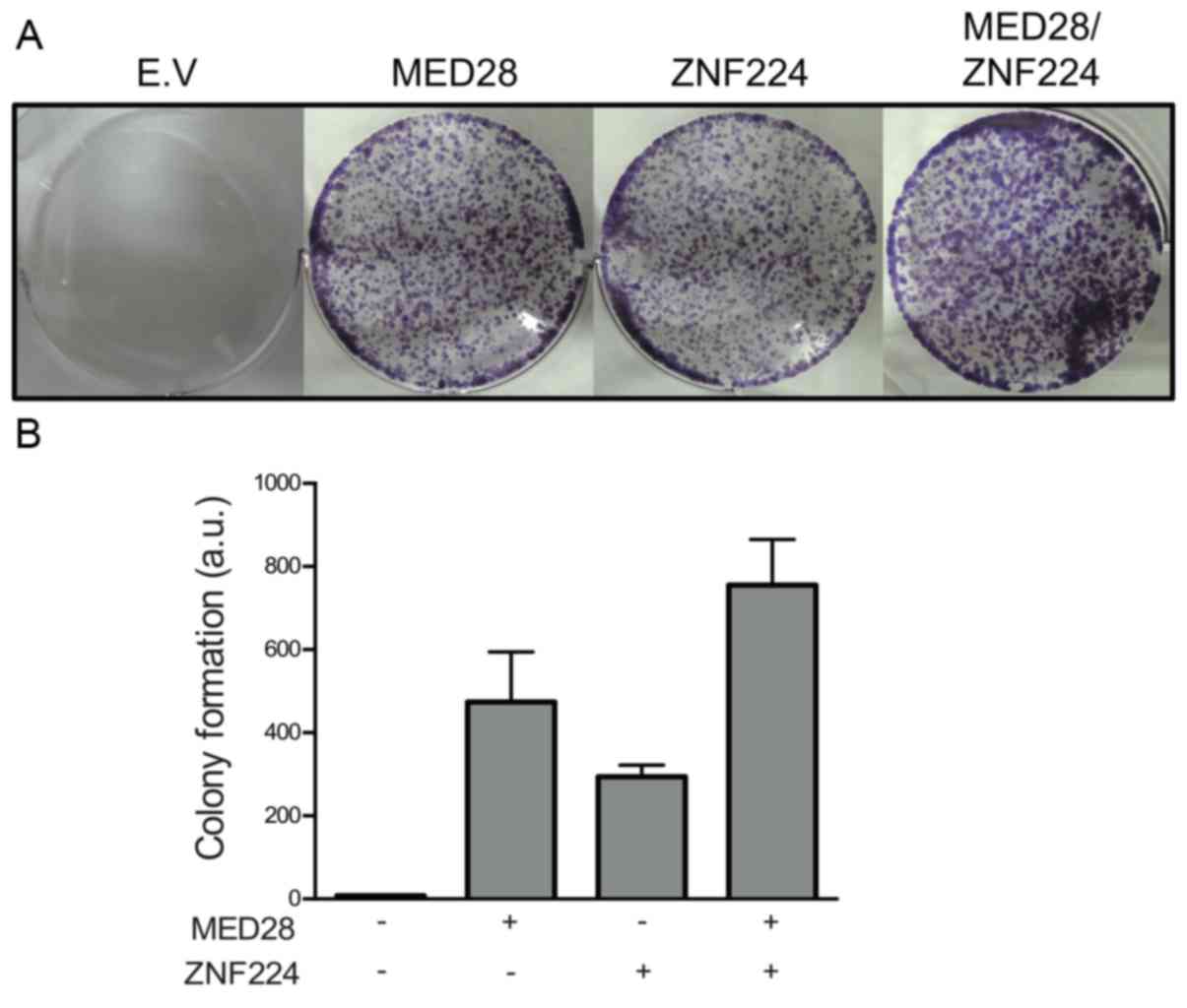
Med28 Increases The Colony Forming Ability Of Breast Cancer Cells By Stabilizing The Znf224 Protein Upon Dna Damage

Figure 4 Cyp24a1 Attenuation Limits Progression Of Brafv600e Induced Papillary Thyroid Cancer Cells And Sensitizes Them To Brafv600e Inhibitor Plx47 Cancer Research

Clonogenic Assay Of Nanoparticles Or Free Dox Or Elc In Hepg2 Cells Download Scientific Diagram


